

Pancreatic Acinar Cell Preparation for Oxygen Consumption and Lactate Production Analysis
用于耗氧量和乳酸生产分析的胰腺腺泡细胞制备
发布: 2020年05月20日第10卷第10期 DOI: 10.21769/BioProtoc.3627 浏览次数: 5002
评审: Khyati Hitesh ShahVanesa Olivares-IllanaWoojong Lee
参见作者原研究论文
The authors used this protocol in:
May 2018
Advertisement


Abstract
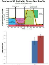
Mitochondrial dysfunction is a principal feature of acute pancreatitis (AP) although the underlying mechanisms are still unclear. AP precipitants induce Ca2+-dependent formation of the mitochondrial permeability transition pore (MPTP) in pancreatic acinar cells (PACs), leading to ATP depletion and necrosis. Evaluations of mitochondrial bioenergetics have mainly been performed in isolated PACs using confocal microscopy, with assessment of mitochondrial membrane potential, NADH/FAD+ and ATP levels, coupled with patch-clamp electrophysiology. These studies are technically demanding and time-consuming. Application of Seahorse flux analysis now allows detailed investigations of bioenergetics changes to be performed in cell populations using a multi-well plate-reader format; rates of oxygen consumption (OCR) and extracellular acidification (ECAR) provide important information about cellular respiration and glycolysis, respectively. Parameters such as maximal respiration, ATP-linked capacity and proton leak can be derived from application of a respiratory function “stress” test that involves pharmacological manipulation of the electron transport chain. The use of Seahorse Flux analysis therefore provides a quick, and convenient means to measure detailed cellular bioenergetics and allows results to be coupled with other plate-reader based assays, providing a fuller understanding of the pathophysiological consequences of mitochondrial bioenergetics alterations.
Keywords: Mitochondrial dysfunction (线粒体功能紊乱)Background
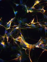
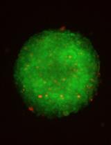
Mitochondrial dysfunction is a core feature of acute pancreatitis (AP), a debilitating and potentially fatal disease for which there is currently no specific therapy (Criddle, 2016; Habtezion et al., 2019). The elucidation of pivotal pathological mechanisms which underlie mitochondrial damage in pancreatic acinar cells (PACs) is paramount for the development of new therapies. Previous evaluations of mitochondrial bioenergetics in isolated PACs have been mostly performed using confocal microscopy, including assessments of mitochondrial membrane potential (tetramethyl rhodamine methyl ester: TMRM), NADH/FAD+ autofluorescence and ATP (Magnesium Green), coupled with patch-clamp electrophysiology (Voronina et al., 2002; Criddle et al., 2004 and 2006). Such studies have pinpointed a reduction of ATP in response to precipitants of AP, via the opening of the mitochondrial permeability transition pore (MPTP), as a critical event that leads to necrotic cell death (Criddle et al., 2006; Mukherjee et al., 2016); supplementation with intracellular ATP ameliorated damage. Furthermore, luciferase measurements in PACs have provided details of changes of both mitochondrial and cytosolic ATP concentrations induced by pathophysiological stimulation (Voronina et al., 2010). Such experimental approaches in single cells, however, are technically difficult and time-consuming. In contrast, population-based assays provide important information about mitochondrial dysfunction and cell death using a convenient plate-reader format (Armstrong et al., 2019). The use of Seahorse Flux analysis allows a detailed evaluation of bioenergetics changes to be performed in PACs, measuring rates of oxygen consumption (OCR) and extracellular acidification (ECAR); these inform about cellular respiration and glycolysis, respectively. A respiratory function “stress” test can further be applied in which pharmacological manipulation of the electron transport chain (ETC) is used to derive parameters such as the Maximal Respiration, Spare Respiratory Capacity, ATP-linked Turnover and Non-mitochondrial Respiration. Such detailed bioenergetics information can be coupled with parallel studies of apoptosis and necrosis to inform the influence of mitochondrial dysfunction on cell death patterns (Armstrong et al., 2018).
Materials and Reagents
- 70 µm filters (Fisherbrand, catalog number: 22363548 )
- Tissue paper (Kimberly-Clark White Roll, Fisher Scientific, catalog number: 12774286 )
- 30 G needle (Microlance 3, 30G x 13 mm) (BD, catalog number: 304000 )
- 1.5 ml microcentrifuge tube (Starlab, catalog number: E1415-1510 )
- Centrifuge tubes
50 ml tube (Starlab, catalog number: E1450-0200 )
15 ml tube (Starlab, catalog number: E1415-0200 ) - Pipette tips
200 µl tip (Starlab, catalog number: S1111-0806 )
1 ml tip (Starlab, catalog number: S1111-6801 ) - Pasteur pipettes 3 ml (Starlab, catalog number: E1414-0311 )
- Male CD1 or C57BL6/J mice, 8-12 weeks old (Charles River)
- Collagenase, Purified (Worthington Biochemical Corporation, catalog number: CLSPA )
- PI-CassetteTM (Chemometec, Nucleocounter, catalog number: 941-0001 )
- Reagent A100; Lysis buffer (SKU: 910-0003, Chemometec, Nucleocounter)
- Reagent B; Stabilizing buffer (SKU: 910-0002, Chemometec, Denmark)
- XF24 Fluxpak, containing cell plates, cartridge consisting of sensor and utility plates and Seahorse XF calibrant (Agilent, Seahorse, catalog number: 100850-100 )
- Matrigel basement membrane matrix (Corning, catalog number: 354234 )
- Dimethyl sulfoxide (DMSO, Sigma, catalog number: D2650 )
- Ethyl alcohol, Pure (Sigma, catalog number: 459836 )
- Oligomycin A (Sigma, Merck, catalog number: 75351 )
- Carbonyl cyanide-4-(trifluoromethoxy)phenylhydrazone (FCCP, C2920, Sigma, Merck)
- Antimycin A from Streptomyces sp. (Sigma, Merck, catalog number: A8674 )
- Rotenone (Sigma, Merck, catalog number: R8875 )
- DMEM (Sigma, Merck, catalog number: D5030 )
- L-Glutamine 100x (Gibco, Life Technologies, cataloge number: 25030-081 )
- D-Glucose (Sigma, Merck, catalog number: G7528 )
- Sodium pyruvate (Sigma, Merck, catalog number: P8574 )
- Sodium chloride (Sigma, Merck, catalog number: S9888 )
- Potassium chloride (Sigma, Merck, catalog number: P3911 )
- Magnesium chloride (Sigma, Merck, catalog number: M1028 )
- Calcium chloride (Sigma, Merck, catalog number: 21115 )
- Extracellular solution (see Recipes)
- Seahorse media (see Recipes)
Equipment
- Surgical scissors
- Water bath
- -20 °C freezer
- Multi-channel pipette 20-200 µl
- Seahorse XF24 Extracellular Flux Analyser (Agilent), although other Seahorse Flux Analysers could be used
- Cell Counter NucleoCounter® NC-100TM (900-0004, ChemoMetec, Denmark)
- Inverted light microscope (GX Microscope XDS-3)
Software
- Wave (Seahorse, Agilent. https://www.agilent.com)
- Prism (GraphPad Software Inc., La Jolla, CA. https://www.graphpad.com)
Procedure
文章信息
版权信息
© 2020 The Authors; exclusive licensee Bio-protocol LLC.
如何引用
Readers should cite both the Bio-protocol article and the original research article where this protocol was used:
- Armstrong, J. A., Sutton, R. and Criddle, D. N. (2020). Pancreatic Acinar Cell Preparation for Oxygen Consumption and Lactate Production Analysis. Bio-protocol 10(10): e3627. DOI: 10.21769/BioProtoc.3627.
- Armstrong, J. A., Cash, N. J., Ouyang, Y., Morton, J. C., Chvanov, M., Latawiec, D., Awais, M., Tepikin, A. V., Sutton, R. and Criddle, D. N. (2018). Oxidative stress alters mitochondrial bioenergetics and modifies pancreatic cell death independently of cyclophilin D, resulting in an apoptosis-to-necrosis shift. J Biol Chem 293(21): 8032-8047.
分类
细胞生物学 > 细胞新陈代谢 > 呼吸测量法
生物化学 > 其它化合物 > 酸
生物化学 > 其它化合物 > 氧
您对这篇实验方法有问题吗?
在此处发布您的问题,我们将邀请本文作者来回答。同时,我们会将您的问题发布到Bio-protocol Exchange,以便寻求社区成员的帮助。