- EN - English
- CN - 中文
Split Nano Luciferase-based Assay to Measure Assembly of Japanese Encephalitis Virus
基于分割纳米荧光素酶的乙型脑炎病毒组装检测
发布: 2020年05月05日第10卷第9期 DOI: 10.21769/BioProtoc.3606 浏览次数: 4322
评审: David PaulAnonymous reviewer(s)
Abstract
Cells infected with flavivirus release various forms of infectious and non-infectious particles as products and by-products. Comprehensive profiling of the released particles by density gradient centrifugation is informative for understanding viral particle assembly. However, it is difficult to detect low-abundance minor particles in such analyses. We developed a method for viral particle analysis that integrates a high-sensitivity split luciferase system and density gradient centrifugation. This protocol enables high-resolution profiling of particles produced by cells expressing Japanese encephalitis virus factors.
Keywords: Flavivirus (黄病毒)Background
The flavivirus, a group of arboviruses including dengue virus, West Nile virus, tick-borne encephalitis virus, Zika virus, yellow fever virus, and Japanese encephalitis virus (JEV), is associated with substantial morbidity and mortality in human populations (Chambers et al., 1990). Flaviviruses have a single ORF genome encoding a long polypeptide, which is post-translationally cleaved into three structural (C, prM and E) and seven non-structural (NS1, NS2A, NS2B, NS3, NS4A, NS4B and NS5) proteins by the host signal peptidase and NS2B-NS3 viral protease (Chambers et al., 1990).
Viral genomic RNA synthesized by the NS5 RNA-dependent RNA polymerase associates with C protein to form the nucleocapsid, which in turn is packaged in the envelope composed of host cell-derived lipid membrane, and viral prM and E transmembrane proteins to form the virion. It has been found that a reduced form of flaviviral genomic RNA, designated as subgenomic replicon, which lacks all structural genes, can replicate itself in host cells (Suzuki et al., 2014), and that the subgenomic RNA is incorporated into the virus-like particle to form single-round infectious particles (SRIPs) when C, prM and E proteins are supplied in trans (Suzuki et al., 2014; Matsuda et al., 2018).
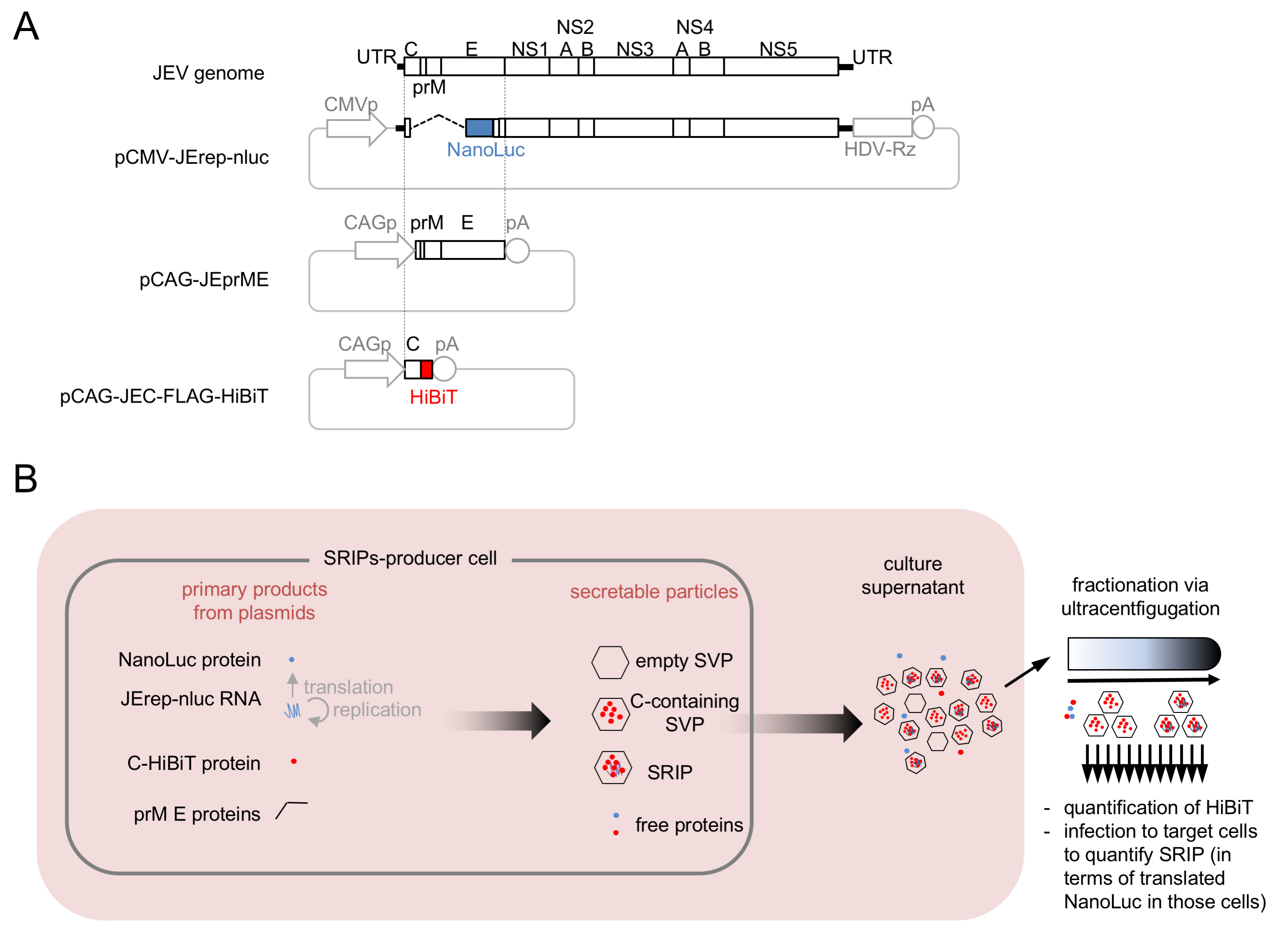
Besides major infectious particles, virus-infected cells often produce minor particles, some of which are hard to detect with immunochemical methods, due to their small amounts. To study typical and atypical particles released from flavivirus-infected cells, we modified a SRIP system (Matsuda et al., 2018) to develop a JEV HiBiT-SRIP assay system. In this system, the C protein is labeled with a highly sensitive HiBiT peptide tag, to enable high-resolution sedimentation profiling of particles produced by the cells expressing viral structural and nonstructural factors (Ishida et al., 2019). Cells transfected with a set of plasmids (referred to as SRIPs-producer cells) produce SRIPs and non-infectious subviral particles containing C-HiBiT, which can be separated by sucrose density gradient centrifugation. SRIPs in sedimentation fractions can be detected by measurement of cellular NanoLuc levels after inoculation of other cells (referred to as target cells) with all the fractions, which allows SRIP-borne NanoLuc expression (Figure 1). It is possible to apply the principle of JEV HiBiT-SRIP assay to other viruses.

Figure 1. Overview of HiBiT-SRIP system. A. HiBiT-SRIP system plasmids. HDV-Rz: self-cleaving HDV Ribozyme; pA: SV40 polyadenylation signal; CMVp: CMV promoter; CAGp: CAG promoter. B. Schematic presentation of HiBiT-SRIP experiments. JErep-nluc: subgenomic replicon with a gene for NanoLuc as the reporter.
Materials and Reagents
- 1.5 ml tube (Watson, catalog number: 131-815C )
- 2 ml tube (Watson, catalog number: 132-620C )
- 6-well cell culture plate (Violamo, catalog number: 2-8588-01 )
- 96-well cell culture plate (Violamo, catalog number: 2-8588-05 )
- 14 x 95 mm open-top polyclear centrifuge tubes (Seton Scientific, catalog number: 7031 )
- White 384-well immuno plates (Thermo, catalog number: 460372 )
- 0.22 μm filter (Merck Millipore, catalog number: UFC30GV00 )
- 293T cells (ATCC, catalog number: CRL-3216 )
- Huh7 cells (JCRB, catalog number: JCRB0403 )
- Plasmid pCAG-JEC-FLAG-HiBiT which encodes HiBiT-tagged wild-type JEV C protein (Ishida et al., 2019; available from authors upon request) or its variant encoding mutant forms of C protein
- Plasmid pCAG-prME which encodes JEV prM and E proteins (Suzuki et al., 2014; available from authors upon request)
- Plasmid pCMV-JErep-nluc which expresses a subgenomic replicon RNA of JEV, which contains a NanoLuc reporter gene (Matsuda et al., 2018, Ishida et al., 2019; available from authors upon request)
- Nano Glo HiBiT lytic detection system (Promega, catalog number: N3040 )
- Nano-Glo luciferase assay system (Promega, catalog number: N1120 )
- Dulbecco’s modified Eagle’s media (Nacalai, catalog number: 08458-16 )
- FBS (fetal bovine serum) (Thermo, catalog number: 10270-106 )
- PBS (phosphate-buffered saline without calcium and magnesium) (Nacalai, catalog number: 14249-24 )
- Benzylpenicillin potassium (Fujifilm, catalog number: 021-07732 )
- Streptomycin sulfate (Tokyo Chemical Industry, catalog number: S0585 )
- PEI MAX (Polyscience, catalog number: 24765-1 ), a transfection reagent
- Opti-MEM (Thermo, catalog number: 31985062 ), reduced serum medium which allows to keep high transfection efficiency with less cytotoxicity
- Sucrose, centrifugal density-gradient grade (Nacalai, catalog number: 30406-25 )
- Tris (Tris (hydroxymethyl) aminomethane) (Nacalai, catalog number: 35406-91 )
- NaCl (Nacalai, catalog number: 31320-05 )
- Triton X-100 (Nacalai, catalog number: 35501-15 )
- KCl (Nacalai, catalog number: 28513-85 )
- Na2HPO4 (Nacalai, catalog number: 31726-05 )
- KH2PO4 (Nacalai, catalog number: 28720-65 )
- PEI solution (1 mg/ml) (see Recipes)
- 100x penicillin G + streptomycin stock solution (see Recipes)
- Culture media (see Recipes)
- 10x PBS (pH 7.4) (see Recipes)
- 10% sucrose/PBS (see Recipes)
- 45% sucrose/PBS (see Recipes)
- 10-45% sucrose linear gradient/PBS (see Recipes)
- Lysis buffer (see Recipes)
Equipment
- Humidified incubator (PHCBI, model: MCO-170AICUVD-PJ , 37 °C, 5% CO2)
- Ultracentrifuge (Hitachi, model: CP80NX )
- Swing bucket rotor (Hitachi, model: P40ST )
- Gradient mixer (BIOCOMP, Gradient Mate)
- Piston gradient fractionator (BIOCOMP, catalog number: 153 )
- Microplate luminometer (Thermo, Varioskan LUX Multimode Microplate Reader)
- -30 °C freezer (PHCBI, catalog number: MDF-MU539D )
Procedure
文章信息
版权信息
© 2020 The Authors; exclusive licensee Bio-protocol LLC.
如何引用
Goto, S., Ishida, K., Suzuki, R. and Morita, E. (2020). Split Nano Luciferase-based Assay to Measure Assembly of Japanese Encephalitis Virus. Bio-protocol 10(9): e3606. DOI: 10.21769/BioProtoc.3606.
分类
分子生物学 > 纳米颗粒 > 植物源纳米颗粒
分子生物学 > 蛋白质 > 蛋白质-蛋白质相互作用
生物化学 > 蛋白质 > 自组装
您对这篇实验方法有问题吗?
在此处发布您的问题,我们将邀请本文作者来回答。同时,我们会将您的问题发布到Bio-protocol Exchange,以便寻求社区成员的帮助。
Share
Bluesky
X
Copy link


