

An ex vivo Approach to Assess Mitochondrial ROS by Flow Cytometry in AAV-tagged Astrocytes in Adult Mice
一种利用流式细胞术进行成年大鼠腺相关病毒标记星型胶质细胞中线粒体ROS水平的体外检测方法
发布: 2020年03月20日第10卷第6期 DOI: 10.21769/BioProtoc.3550 浏览次数: 6590
评审: Arnau Busquets-GarciaEtienne Hebert-ChatelainAnonymous reviewer(s)
参见作者原研究论文
The authors used this protocol in:

Feb 2019


Abstract
Mitochondrial reactive oxygen species (mROS) are naturally produced signalling molecules extremely relevant for understanding both health- and disease-associated biological processes. The study of mROS in the brain is currently underway to decipher their physiopathological roles and contributions in neurological diseases. Recent advances in this field have highlighted the importance of studying mROS signalling and redox biology at the cellular level. Neurons are especially sensitive to the harmful effects of excess mROS while astrocytic mROS have been shown to play a relevant physiological role in cerebral homeostasis and behaviour. However, given the complexity of the brain, investigating mROS formation in a specific cell-type in adult animals is methodologically challenging. Here we propose an approach to specifically assess mROS abundance in astrocytes that combines i) a targeting strategy based on the use of adeno-associated virus (AAV) vectors expressing the green fluorescent protein (GFP) under an astrocyte (glial fibrillary acidic protein or GFAP) promoter, along with, ii) a robust and widely extended protocol for the measurement of mROS by flow cytometry using commercial probes. The significance of this work is that it allows the selective study of astrocytic mROS abundance by means of easily accessible technology.
Keywords: Reactive Oxygen Species (ROS) (活性氧)Background
Oxidative damage is associated with the aetiology of many diseases, including neurodegenerative disorders given that the brain is an exceptionally vulnerable tissue to oxidative stress as well as to age-related alterations (Cobley et al., 2018; Mattson and Arumugam, 2018). Yet there are still important gaps in the understanding of reactive oxygen species (ROS) pathophysiology in the brain. The deleterious effects associated with ROS in situations of redox stress are in contrast to the increasing evidence suggesting that physiological processes are also fine-tuned by ROS (D’Autréaux and Toledano, 2007; Holmström and Finkel, 2014; Hopkins, 2016a and 2016b). In fact, the clinical application of ROS as signalling molecules is still far away from being an effective solution as a translational therapy (Kamat et al., 2008; Carvalho et al., 2017). Thus, the success of antioxidant therapies depends on considering aspects of redox biology such as its pleiotropism or its cellular and subcellular origin (Juránek et al., 2013; Zhang et al., 2016). In vitro studies using brain cells, principally neurons and astrocytes, have been essential for understanding the highly distinctive biology of these cells. In this sense, it has been shown how these brain cells present different molecular specialization, highlighting the importance of crosstalk during neuron-astrocyte coupling which ensures brain bioenergetic and redox homeostasis (Fernandez‑Fernandez et al., 2012; Bolaños, 2016). Regarding the study of ROS in brain cells, there is recent evidence indicating that the levels of mitochondrial reactive oxygen species (mROS) are immensely greater–about one order of magnitude–in astrocytes than in neurons (Lopez-Fabuel et al., 2016). This finding reiterates the relevance of considering the cellular origin of neural ROS as an important new factor in the study of redox biology in the brain.
To address this issue, we have reported the use of a new method to quantify the levels of mROS in adult mice astrocytes ex vivo, independently of neurons (Vicente-Gutierrez et al., 2019). This methodology has allowed us to demonstrate an effective and specific down-modulation of astrocytic mROS in a transgenic mouse model expressing a mitochondrial-tagged form of catalase (mitoCAT or mCAT) (Vicente-Gutierrez et al., 2019). In this particular study, we showed that astrocytic mROS has an impact on neuronal function and survival by regulating bioenergetics and redox metabolism. Thus, by decreasing endogenous levels of astrocytic mROS, we found that astrocytic mROS may modulate glucose utilization and neuronal function in behaving mice (Vicente-Gutierrez et al., 2019). Using the methodology that we herein describe, we postulated that endogenous mROS in astrocytes play a physiological role in the maintenance of brain homeostasis. Furthermore, in this same study (Vicente-Gutierrez et al., 2019), we were able to characterize a novel conditional mCAT mouse that could be useful for testing the implications of mROS in a desired specific tissue type or pathological model.
Since the study of ROS depends not only on concentration but also on its spatiotemporal distribution, real-time imaging of ROS, possibly in vivo, has become necessary in order for scientists to determine which of their biological activities may present a potential for clinical translation. However, this objective is still unachievable owing to current available techniques (Maulucci et al., 2016). Here, we describe in further detail the protocol previously used in Vicente-Gutierrez et al. (2019) to measure mROS abundance, specifically superoxide anion-abundance in astrocytes ex vivo. Although there are different probes commercially available, the most widely used is MitoSOXTM. The MitoSOX Red mitochondrial superoxide indicator enters into mitochondria, where it accumulates in response to the mitochondrial membrane potential (ΔΨm) and becomes oxidized. This probe has been widely used in astrocytes in culture (Sheng et al., 2013; Angelova et al., 2015) and in cultured hippocampal sections (Ishii et al., 2017). MitoSOX fluorescence intensity is commonly assessed by microscope imaging or flow cytometry. As mentioned above, this fluorescent dye requires an active mitochondrial membrane potential to enter into mitochondria. This implies that to measure mROS, it is necessary to use live cells and to determine, in parallel, the ΔΨm in order to confirm that differences in the MitoSOX signal are independent of ΔΨm. However, this fact presents a challenge when working with fixed-cells. For instance, this exclude the use of imaging techniques like immunohistofluorescence in fixed-cells which, in addition, to study individual cellular phenomena required the use of different cell markers. To overcome the cell origin problem, others have used genetically encoded probes to assess redox histology in mouse (Fujikawa et al., 2016). Thus, immunohistochemical strategies still offer a suitable approach for detecting the footprints of redox stress. Nowadays, two-photon laser scanning microscopy has become an interesting tool for studying cellular parameters in awake mice, in vivo. However, this technology is not easily accessible in every laboratory and is limited to studying superficial brain areas (Pérez-Alvarez et al., 2013; Wang et al., 2017). Another drawback to this technique is the large amount of time required for sample preparation and for obtaining replicas. In contrast, the protocol that we will describe is relatively easy, accessible, fast and robust.
The ability of certain adeno-associated virus (AAV) vectors to cross the blood-brain barrier after intravenous injection makes it possible to obtain transgene expression in brain cells. The AAV-PHP.eB capsid is one of the serotypes that is able to efficiently transduce the central nervous system (Chan et al., 2017) and is useful for many applications. For instance, we have used it to express the green fluorescent protein (GFP) under the control of the astrocyte-specific glial fibrillary acidic protein (GFAP) short promoter (gfa-ABC1D) (Lee et al., 2008). Astrocytes were specifically tagged in vivo by infecting mice intravenously through the retro-orbital venous sinus with the AAV-PHP.eB-gfa-ABC1D-GFP construct. Then, we obtained a single brain cell suspension and adapted a well-known protocol to measure MitoSOX by flow cytometry. Therefore, this protocol combines the use of accessible technology like flow cytometry with the use of commercial probes. Accordingly, another advantage to this approach is its versatility, since it is also useful for assessing other cell-specific processes in brain function. By using other brain cell-specific promoters to target neurons, microglia or oligodendrocytes, for instance, the same phenomenon could be measured independently or simultaneously in different cell types. Moreover, this protocol allows the same sample to be analysed using different commercially available probes to measure different biological processes. Finally, this tool is greatly useful to study other cellular and subcellular phenomena and can be used at different time points during model lifespan as well as different tissue types. Overall, we believe that this straight-forward protocol for measuring mROS in adult astrocytes is beneficial for studying redox biology in vivo at the cellular level in the brain.
Materials and Reagents
- Anaesthesia
- Mice C57BL/6J
- Sevofluorane (Sevorane®)
- Oxygen (O2) Purity: 99.995% (Air Liquide, AlphagazTM)
- Nitrous oxide (N2O) (Air Liquide, AlphagazTM, CAS no.: 010024-97-2)
- AAVs injection
- High level surface disinfectant Rely+OnTM Virkon® (DuPont®)
- Barrier and non-filtered pipette Tips (Art tips, Thermo Scientific)
- Insulin syringes BD Micro-Fine + Demi 0.3 ml, 30 Gauge, 8 mm (Becton Dickinson, catalog number: 324826 )
- AAVs. To target astrocytes in this protocol we use AAV-PHP.eB-gfa-ABC1D-GFP construct
- Pluronic F-127 (Sigma-Aldrich, catalog number: P2443-2506 )
- Phosphate buffered saline (PBS) (GibcoTM, catalog number: 70011044 )
- Parafilm paper (Bemis, PM-996, USA)
- Brain single-cell suspension
- Microsurgical scissor (Fine Science Tools, catalog number: 91501-09 )
- Scalpel blade (N° 24, KRAPE S.A., catalog number: BS EN 27740 )
- Fetal bovine serum (FBS) (GibcoTM, catalog number: 10270 )
- Bovine serum albumin (Sigma-Aldrich)
- DNase I (Roche Diagnostics, catalog number: 10104159001 )
- Trypsin (Sigma-Aldrich, catalog number: T4799 )
- NaCl (Merck, catalog number: 106404 )
- KCl (Merck, catalog number: 104936 )
- MgSO4·7H2O (Merck, catalog number: 5886 )
- NaHCO3 (Sigma, catalog number: 6329 )
- NaH2PO4·2H2O (Merck, catalog number: 106345 )
- β-D(+)Glucose (Sigma, catalog number: G5250 )
- Phenol red. (Sigma, catalog number: P5530 )
- KH2PO4 (Merck, catalog number: 4873 )
- HEPES (Sigma, catalog number: H3375 )
- CaCl2·2H2O (Merck, catalog number: 102381 )
- Earle’s Balanced Salt Solution (EBSS) buffer (see Recipes)
- HBSS buffer (see Recipes)
- Disaggregation solution (see Recipes)
- Resuspension solution (see Recipes)
- Mitochondrial ROS measurement
- MitoSOXTM Red Mitochondrial Superoxide Indicator (Molecular Probes, Inc., InvitrogenTM, Thermo Fisher, catalog number: M36008 )
Equipment
- Anaesthesia procedure and AAVs handling
- Automatic pipettes (Gilson's PIPETMANTM)
- Microcentrifuge (Eppendorf, model: Centrifuge 5424 )
- Vortex (Scientific Industries, Inc., model: Vortex Genie 2 )
- Anaesthesia system composed by a gas distribution column (Hersill H-3, Spain) and a Vaporizer (InterMed Penlons Sigma Delta) (Figure 1)
- Class II cabinet equivalent to Telstar Bio II Advance certified according EN-12469-2000
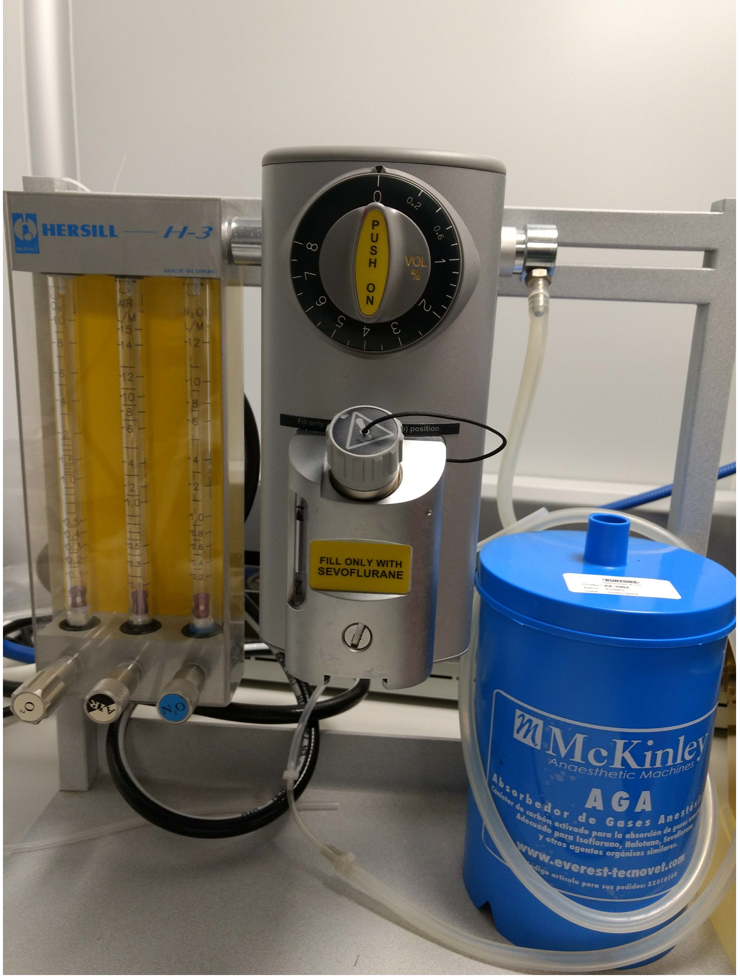
Figure 1. Anaesthesia system
- Mitochondrial ROS measurement
- FACSCaliburTM Flow Cytometer (Becton Dickinson, catalog number: 342975 ), equipped with a15 mW argon laser
- Centrifuge adapted for Flow cytometry tubes (Eppendorf, model: Centrifuge 5810 R )
Software
- BD CellQuestTM Pro version 5.2 (Becton, Dickinson & Company, BD Biosciences)
- FlowJo X 10.0.7r2 (FlowJo, LLC, Becton, Dickinson & Company)
Procedure
文章信息
版权信息
© 2020 The Authors; exclusive licensee Bio-protocol LLC.
如何引用
Vicente-Gutierrez, C. and Bolaños, J. P. (2020). An ex vivo Approach to Assess Mitochondrial ROS by Flow Cytometry in AAV-tagged Astrocytes in Adult Mice. Bio-protocol 10(6): e3550. DOI: 10.21769/BioProtoc.3550.
分类
神经科学 > 细胞机理 > 线粒体
神经科学 > 神经系统疾病 > 细胞机制
细胞生物学 > 细胞信号传导 > 胞内信号传导
您对这篇实验方法有问题吗?
在此处发布您的问题,我们将邀请本文作者来回答。同时,我们会将您的问题发布到Bio-protocol Exchange,以便寻求社区成员的帮助。