

Mouse Embryonic Tooth Germ Dissection and Ex vivo Culture Protocol
小鼠胚胎牙胚分离和体外培养方案

(*contributed equally to this work) 发布: 2020年02月05日第10卷第3期 DOI: 10.21769/BioProtoc.3515 浏览次数: 6853
评审: Ralph Thomas BoettcherIrit AdiniThirupugal Govindarajan
参见作者原研究论文
The authors used this protocol in:
Sep 2018


Abstract
A tooth germ ex vivo organ culture allows visualization of its development in different stages, thus enabling investigation of the molecular mechanisms of regulatory factors. Tooth germs can be rapidly dissected from E13 mouse embryos and placed on cell culture inserts for observation of subsequent tooth germ development in a three-dimensional situation in real time. This method is also suitable for other organs that develop by epithelial-mesenchymal interactions, including salivary gland, hair, lung, and kidney. In addition, siRNAs or growth factors can be easily added to ex vivo tooth germ cultures to investigate the detailed molecular function of specific genes. The present protocol provides an efficient and practical method for isolation and ex vivo culture of embryonic tooth germs.
Keywords: Organ culture (器官培养)Background
Teeth are considered to represent a typical epithelial-mesenchymal organ. When investigating tooth development, observation of the developmental pattern can help to elucidate the mechanisms of morphogenesis biology. When transfecting siRNAs or expression vectors into embryonic organs, a maternal injection protocol is commonly used in studies of mice. However, with traditional methods, it is difficult to efficiently transport reagents into the target organ. On the other hand, an ex vivo organ culture simplifies direct transportation of reagents into a dissected organ, allowing for observations in a three-dimensional manner. In addition to teeth, other epithelial-mesenchymal organs such as salivary gland and hair can also be successfully cultured with this system.
Several different organ culture protocols have been presented for tooth germ organ culturing, such as a semi-solid method (Heywood and Appleton, 1984), with Matrigel (Shih and Sander, 2014; Sun et al., 2016), and with the use of mesh placed in cell culture dishes (Yamada et al., 1980; Bringas et al., 1987; Tabata et al., 1996; Fukumoto et al., 2006; Yamamoto et al., 2008; Yamada et al., 2016). However, each requires preparation of the culture materials in the wells every time prior to organ dissection. In contrast, with the present organ culture system, the materials can be directly purchased from a supplier, which saves time and avoids inevitable contamination during preparation of the culture materials. Traditional methods used to examine the functions of gene or drugs in vivo require time to determine the results individually, whereas with an ex vivo culture, it is possible to examine a large number of organ samples at the same time. Furthermore, it is possible to screen for critical genes or other related factors during organ development.
Here, we describe a relatively simple and efficient method for ex vivo organ culture of embryonic tooth germs. With the use of this protocol, screening studies and development observations can be objectively and directly conducted.
Materials and Reagents
- 100 mm cell culture dish (Falcon, catalog number: 353003)
- 60 mm cell culture dish (Falcon, catalog number: 353002)
- Cell culture 6-well plate for insert (Falcon, catalog number: 353502)
- Cell culture insert (6 wells, 8 μm, Falcon, catalog number: 353093)
- Thirteen-day-pregnant female mouse
- Fetal bovine serum (FBS, Gibco, catalog number: 10437028), short term storage at 4 °C, long term storage at -20 °C, heat inactivation not necessary
- siRNA: Nkx2-3 (Dharmacon, ON-TARGET Plus L-057189-01-0002), storage at -20 °C; control (Dharmacon, ON- TARGET Plus Nontargeting Control Pool D-001810-05), storage at -20 °C
- 70% ethanol
- Dulbecco’s phosphate buffered saline without Ca2+ or Mg2+ (D-PBS(-), Wako, catalog number: 04529795), storage at room temperature
- Dulbecco’s modified Eagle’s medium (DMEM)/F-12 (Gibco, catalog number: 11330032), storage at 4 °C
- L-Glutamine (200 mM, 100x) (Gibco, catalog number: 25030081), storage at -20 °C
- Penicillin-streptomycin (Liquid, Gibco, catalog number: 15140122), storage at -20 °C
- L-Ascorbic acid (Sigma-Aldrich, catalog number: A4544-25G), storage at room temperature
- Lipofectamine 3000 with Plus reagent (Life Technologies, catalog number: L3000-015), storage at 4 °C
- Opti-MEM (Gibco, catalog number: 31985062), storage at 4 °C
- Working medium (see Recipes)
- Organ culture medium (see Recipes)
- siRNA transfection medium (see Recipes)
Equipment
- Surgical instruments: tweezers, scissors, fine scissors, precision tweezers (Dumont #5 Forceps) (Figure 1A)
- Compact Greenough Stereo Microscope (ZEISS, model: Stemi 305 EDU) (Figure 1B)
- Mouse dissection table
- CO2 incubator (ASTEC, model: SCA-80DS)
- Research inverted system microscope (Olympus, model: IX71)
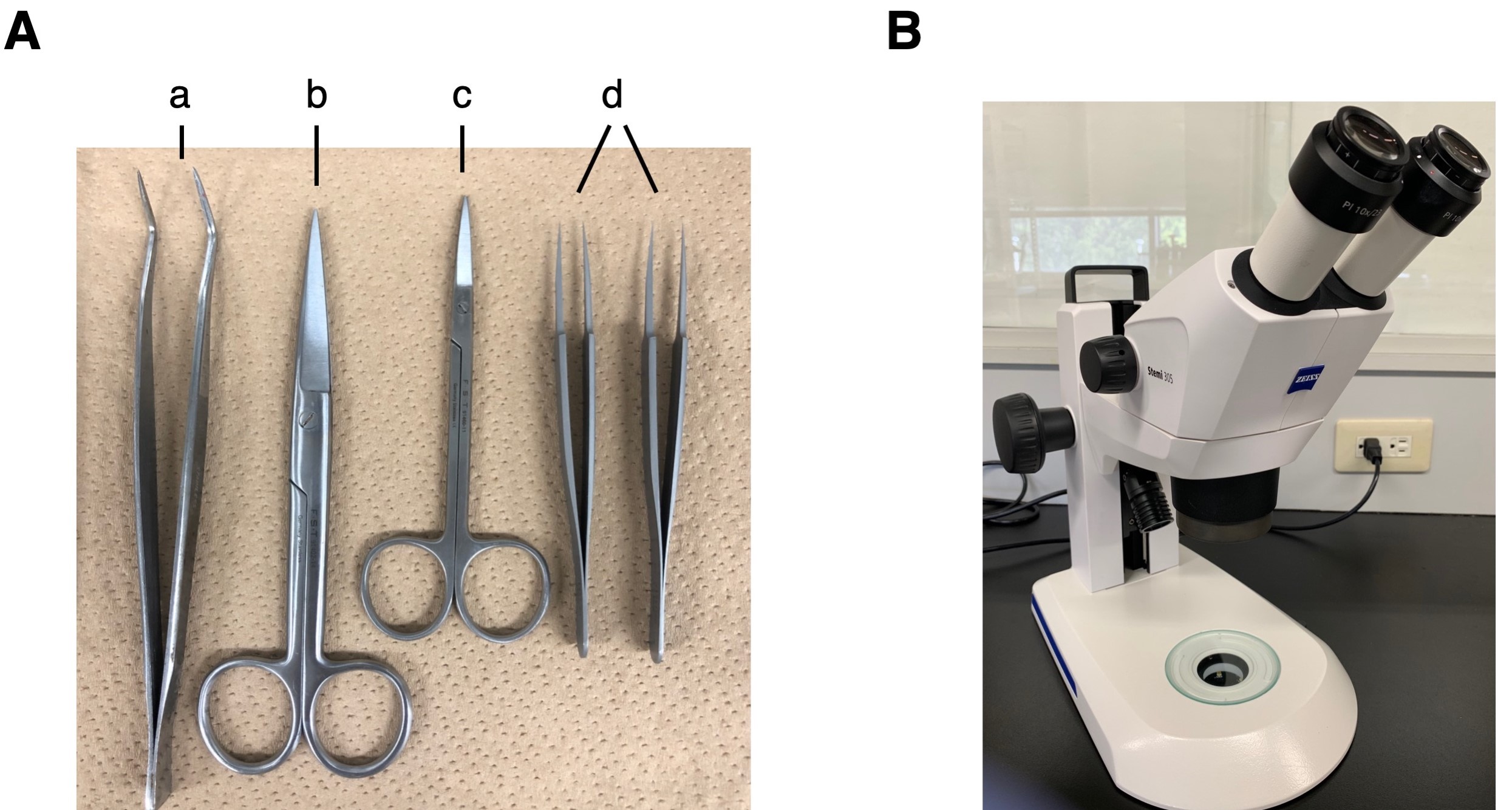
Figure 1. Instruments used for embryonic mandible isolation and molar germ dissection. A. Surgical instruments: a. tweezers, b. scissors, c. fine scissors, d. precision tweezers. B. Compact Greenough stereo microscope.
Procedure
文章信息
版权信息
© 2020 The Authors; exclusive licensee Bio-protocol LLC.
如何引用
Readers should cite both the Bio-protocol article and the original research article where this protocol was used:
- Han, X., Yoshizaki, K., Tian, T., Miyazaki, K., Takahashi, I. and Fukumoto, S. (2020). Mouse Embryonic Tooth Germ Dissection and Ex vivo Culture Protocol. Bio-protocol 10(3): e3515. DOI: 10.21769/BioProtoc.3515.
- Han, X., Yoshizaki, K., Miyazaki, K., Arai, C., Funada, K., Yuta, T., Tian, T., Chiba, Y., Saito, K., Iwamoto, T., Yamada, A., Takahashi, I. and Fukumoto, S. (2018). The transcription factor NKX2-3 mediates p21 expression and ectodysplasin-A signaling in the enamel knot for cusp formation in tooth development. J Biol Chem 293(38): 14572-14584.
分类
发育生物学 > 形态建成 > 器官形成
细胞生物学 > 细胞分离和培养 > 细胞分离
细胞生物学 > 细胞分离和培养 > 器官培养
您对这篇实验方法有问题吗?
在此处发布您的问题,我们将邀请本文作者来回答。同时,我们会将您的问题发布到Bio-protocol Exchange,以便寻求社区成员的帮助。