

Detection of Individual RNA in Fixed Cells and Tissues by Chromogenic ISH
利用显色原位杂交进行固定细胞和组织中单个RNA的检测
发布: 2020年02月05日第10卷第3期 DOI: 10.21769/BioProtoc.3510 浏览次数: 5230
评审: Imre GáspárNidhi SharmaShalini Low-Nam
参见作者原研究论文
The authors used this protocol in:
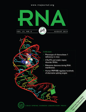
Aug 2019
Advertisement


Abstract
Visualization of RNA molecules in situ helps to better understand the functions of expressed genes. Currently, most conventional in situ hybridization methods for visualization of individual RNAs are based on fluorescence detection. Herein we present a chromogenic in situ hybridization protocol for visualization of single RNA molecules in fixed cells and tissues. The protocol is based on padlock probing and rolling circle amplification to generate detectable chromogenic signal from single RNA molecules. Chromogenic signal can avoid background autofluorescence and can be preserved for a longer period than fluorescence signal.
Keywords: Chromogenic in situ hybridization (显色原位杂交)Background
The abundance and spatial location of expressed RNA molecules indicates the physiological and pathological status of cells and tissues. Therefore, there is an increasing interest for in situ RNA detection. Compared to conventional in situ hybridization methods, novel methods that can detect RNA at individual molecule level are more sensitive and specific (Crosetto et al., 2015). These methods include single molecule fluorescence in situ hybridization (smFISH) (Femino et al., 1998), methods based on rolling circle amplification (Larsson et al., 2010), hybridization chain reaction (HCR) (Shah et al., 2016), and branched DNA (bDNA) technology (Wang et al., 2012; Battich et al., 2013). Current methods for individual RNA in situ detection assays are normally readout as fluorescence signal, which offers good sensitivity and is easy to multiplex. Chromogenic readout is often used in conventional RNA ISH assays, providing stable signal that can be stored for long period of time, and is not affected by autofluorescence or photobleaching. Therefore, we introduce the single molecule chromogenic in situ hybridization (smCISH) assay that enables sensitive and specific detection of individual RNA in fixed cells and tissues (Jiang et al., 2019). Our protocol is based on padlock probing and rolling circle amplification (Nilsson et al., 1994; Banér et al., 1998). First, padlock probe is designed to directly bind to target RNA specifically. After padlock probe hybridization, probe circularization is performed using SplintR DNA ligase. The closed circle is RNA-templated ligation of its two ends. Next, an RCA primer is hybridized to the circularized padlock probe to initiate rolling circle amplification reaction. The padlock probe is then amplified into its complementary concatenated form to generate RCP. HRP labeled detection probes are then hybridized to the RCP. Finally, chromogenic signal is developed using H2O2 and 3,3'-Diaminobenzidine (DAB) to generate brown insoluble dots from individual RCPs, which corresponds to single RNA molecules. Our results show that single molecule chromogenic in situ hybridization assay can count and localize individual RNA molecules in fixed cells and tissue samples.
Materials and Reagents
- Secure-Seal hybridization chamber, 9 mm diameter, 0.8 mm deep (Containing seal tabs, Thermo Scientific, catalog number: S24732)
- EasYFlask 25 cm2 (Thermo Scientific Nunc, catalog number: 156340-ROS)
- Serological Pipette (Thermo Scientific Nunc, 2 ml, 5 ml, 10 ml, 50 ml)
- Cell culture Dish, 150 mm x 20 mm (Thermo Scientific Nunc, catalog number: 168381-ROS)
- Adhesion Microscope Slides (Citotest, catalog number: 188105)
- 15 ml Conical Centrifuge Tubes (Thermo Scientific Nunc, catalog number: 339650-SPL)
- 50 ml Conical Centrifuge Tubes (Thermo Scientific Nunc, catalog number: 339652-SPL)
- Filtered Pipette Tip (QSP, 0.1-10 µl, 2-20 µl, 10-100 µl, 20-200 µl, 100-1,000 µl)
- 0.2 ml PCR Tubes (AXYGEN, catalog number: PCR-02-C)
- 1.5 ml Microtubes (AXYGEN, catalog number: MCT-150-C)
- 0.22 μm membrane filter (Millipore, catalog number: SLGP033RS)
- HER2 padlock probe (sequence: 5′-ATTACTTGCAGGTTCTTTCCTTTTACGACCTCAATGCTGCTGCTGTACTACTCTTCAGAATTCGTCCCCGG, underline: target hybridization sequences andd detection probe complementary part, bold italics: RCA primer binding part) (Synbio Tech, China)
- ACTB padlock probe (sequence: 5′-CTGTGCTCGCGGGGCGTTCCTTTTACGACCTCAATGCTGCTGCTGTACTACTCTTGGCAAAGGCGAGGCT, underline: target hybridization sequences and detection probe complementary part, bold italics: RCA primer binding part) (Synbio Tech, China)
- RCA primer (sequence: 5′-ACAGCAGCAGCATTGAGGTC) (Synbio Tech, China)
- HRP labeled detection probe (sequence: 5′ HRP-CCTCAATGCTGCTGCTGTACTAC) (TaKaRa)
- Pepsin (Sigma-Aldrich, catalog number: P7012-1G), store at -20 °C
- Fetal Bovine Serum (Hyclone, catalog number: 10270-106), store at -20 °C
- Trypsin (Hyclone, catalog number: SH30042.01), store at -20 °C
- ATP, 100 mM Solution (Thermo Scientific, catalog number: R0441), store at -20 °C
- T4 Polynucleotide Kinase, 10 U/µl (Thermo Scientific, catalog number: EK0032, containing PNK buffer A), store at -20 °C
- dNTP Set, 100 mM Solutions (Thermo Scientific, catalog number: R0182), store at -20 °C
- SplintR Ligase, 25 U/µl (NEB, catalog number: M0375L), store at -20 °C
- phi29 DNA Polymerase, 10 U/µl (Thermo Scientific, catalog number: EP0094), store at -20 °C
- BSA, 20 mg/ml (NEB, catalog number: B9000S), store at -20 °C
- RiboLock RNase Inhibitor, 40 U/µl (Thermo Scientific, catalog number: EO0384), store at -20 °C
- Tween-20 (Sigma-Aldrich, catalog number: P9416-100ML), store at room temperature (RT)
- Triton X-100 (Sigma-Aldrich, catalog number: T8787-100ML), store at RT
- NaOH (Sigma-Aldrich, catalog number: 795429-500G), store at RT
- KCl (Sigma-Aldrich, catalog number: P9541-500G), store at RT
- NaCl (Sigma-Aldrich, catalog number: S3014-1kG), store at RT
- Na2HPO4 (BBI LIFE SCIENCES, catalog number: A610404-0500), store at RT
- KH2PO4 (Sangon Biotech, catalog number: A100781-0500), store at RT
- Sodium iodate (BBI LIFE SCIENCES, catalog number: A600859), store at RT
- Aluminum potassium sulfate dodecahydrate (Sangon Biotech, catalog number: A500755), store at RT
- Glycerol (Sigma-Aldrich, catalog number: G9012-100ML), store at RT
- Hematoxylin (Sigma-Aldrich, catalog number: H3136-25G), store at RT
- Neutral balsam mounting medium (BBI LIFE SCIENCES, catalog number: E675007), store at RT
- Xylene (XILONG SCIENTIFIC, catalog number: 33535-500ML), store at RT
- Ethanol (Macklin, catalog number: E809061-500 ML), store at RT
- Acetate (Macklin, catalog number: A801295-500 ML), store at RT
- Hydrochloric acid (SCR, catalog number: 10011018), store at RT
- DEPC (Sigma-Aldrich, catalog number: D5758-25ML), store at 4 °C
- Paraformaldehyde (Sigma-Aldrich, catalog number: 16005-1KG-R), store at 4 °C
- Formamide (Sigma-Aldrich, catalog number: F9037-100ML), store at 4 °C
- 20x SSC buffer (Sigma-Aldrich, catalog number: S6639), store at 4 °C
- 1x Phosphate Buffered Saline (Hyclone, catalog number: SH30256.01), store at 4 °C
- DNase/RNase-Free Water (Solarbio, catalog number: R1600), store at 4 °C
- Hydrogen peroxide solution, 30 wt.% in H2O (Macklin, catalog number: H811240-500 ml), store at 4 °C
- DAB Immunohistochemistry Color Development Kit (BBI LIFE SCIENCES, catalog number: EP670033), store at -20 °C
- EDTA (Sigma-Aldrich, catalog number: RDD017-500G), store at RT
- Serum-free medium, DMEM/HIGH GLUCOSE (Hyclone, catalog number: SH30022.01), store at 4 °C
- Penicillin-Streptomycin (Hyclone, catalog number: SV30010), store at -20 °C
- Culture media (50 ml) (see Recipes)
- 10x PBS (500 ml), pH 6.8 (see Recipes)
- 1x DEPC-PBS (1 L), pH 7.4 (see Recipes)
- 1x DEPC-PBST (1 L) (see Recipes)
- 4% (w/v) PFA (500 ml) (see Recipes)
- 1x TE buffer (100 ml), pH 8.0 (see Recipes)
- 0.5%(v/v) Triton X-100 (5 ml) (see Recipes)
- 3% (w/v) H2O2 solution (50 μl) (see Recipes)
- 2x hybridization buffer (5 ml) (see Recipes)
- Washing buffer (15 ml) (see Recipes)
- 2x detection buffer (5 ml) (see Recipes)
- Staining buffer (50 μl) (see Recipes)
- 0.1% hydrochloric acid-ethanol (5 ml) (see Recipes)
- Hematoxylin Staining Solution (50 ml) (see Recipes)
- 25 mM dNTPs (100 μl) (see Recipes)
Equipment
- Vortex Mixers (MIULAB, model: MIX-25P)
- Microcentrifuge (MIULAB, model: Mini-6K)
- Magnetic stirrer (IKA, RCT basic)
- pH meter (OHAUS, model: starter3100)
- PCR machine (BIOER, model: TC-XP)
- Analytical balance (OHAUS, model: AS220.R2)
- Pipette (Eppendorf AG, Research plus)
- Cell culture incubator (Esco, model: CCL-170B-8)
- Biosafety cabinet (Esco, model: AC2-4S1)
- Ultra-low temperature freezer (Thermo, model: DW-86L338)
- Freezer (Siemens, model: KG32EV2S0C)
- Electro-heating standing-temperature cultivator (ENXIN, model: DRP-9052)
- Microscope (Leica, model: DM6B, equipped with a DFC7000T camera)
Software
- CellProfiler
- GraphPad Prism
Procedure
文章信息
版权信息
© 2020 The Authors; exclusive licensee Bio-protocol LLC.
如何引用
Jiang, M., Lin, C. and Ke, R. (2020). Detection of Individual RNA in Fixed Cells and Tissues by Chromogenic ISH. Bio-protocol 10(3): e3510. DOI: 10.21769/BioProtoc.3510.
分类
癌症生物学 > 通用技术 > 生物化学试验 > RNA
细胞生物学 > 细胞成像 > 共聚焦显微镜
分子生物学 > RNA > RNA 标记
您对这篇实验方法有问题吗?
在此处发布您的问题,我们将邀请本文作者来回答。同时,我们会将您的问题发布到Bio-protocol Exchange,以便寻求社区成员的帮助。