

Visualization of Actin Organization and Quantification in Fixed Arabidopsis Pollen Grains and Tubes
固定拟南芥花粉粒及花粉管中肌动蛋白组织可视化及定量分析


发布: 2020年01月05日第10卷第1期 DOI: 10.21769/BioProtoc.3509 浏览次数: 5391
评审: Zhibing LaiYing FengSam-Geun Kong
参见作者原研究论文
The authors used this protocol in:

Dec 2015
Advertisement


Abstract
Although it is widely accepted that actin plays an important role in regulating pollen germination and pollen tube growth, how actin exactly performs functions remains incompletely understood. As the function of actin is dictated by its spatial organization, it is the key to reveal how exactly actin distributes in space in pollen cells. Here we describe the protocol of revealing and quantifying the spatial organization of actin using fluorescent phalloidin-staining in fixed Arabidopsis pollen grains and pollen tubes. We also introduce the method of assessing the stability and/or turnover rate of actin filaments in pollen cells using the treatment of latrunculin B.
Keywords: Arabidopsis (拟南芥)Background
Pollen germination and subsequent pollen tube growth is the critical step during sexual plant reproduction, as it provides passage for the delivery of two non-motitle sperm cells to finally effect double fertilization in flowering plants (McCormick, 2013). How pollen germination and pollen tube growth are regulated has been subject to intensive scrutiny in the past several decades. It was shown that actin is an essential regulator of pollen germination and pollen tube growth and morphogenesis (Gibbon et al., 1999; Vidali et al., 2001). As pollen germination and pollen tube growth are very sensitive to the perturbation on the actin cytoskeleton (Gibbon et al., 1999; Vidali et al., 2001), and considering along with the fact that Arabidopsis thaliana is amenable to genetic analysis, Arabidopsis pollen has increasingly becoming the great cellular system in documenting the organization and regulation of the actin cytoskeleton toward understanding how actin performs functions in pollen (Qu et al., 2015). Arabidopsis pollen cell is an excellent cellular system to study the organization and regulation of the actin cytoskeleton is also because the spatial organization of actin is very regular in pollen cells (Ren and Xiang, 2007; Cheung and Wu, 2008; Chen et al., 2009; Staiger et al., 2010; Fu, 2015; Qu et al., 2015), and it is therefore comparatively easy to quantify the organization of actin in pollen cells (Fu, 2015; Qu et al., 2015). The colleagues in this field have developed different methods in revealing the organization of actin in pollen cells, including staining with fluorescent-phalloidin or immune-staining probed with anti-actin antibody in fixed Arabidopsis pollen cells as well as decoration with different actin markers in living pollen cells. Compared to immuno-staining with anti-actin antibody in fixed pollen cells, actin staining with fluorescent phalloidin can yield more cleanly filamentous images as phalloidin specifically binds to actin filaments with high affinity that reduces the background noise, which will facilitate subsequent analysis and quantification of actin organization in pollen cells. In addition, the procedure of actin staining with fluorescent phalloidin is comparatively simple and straightforward. Furthermore, the whole procedure was completed on solid pollen germination medium, which minimizes the effect of various treatments on the morphology of pollen tubes. This method has been carried out routinely and successfully in our laboratory. The detailed method can be found in series of our previously published papers (Wu et al., 2010; Zhang et al., 2010; Qu et al., 2013; Zheng et al., 2013; Chang and Huang, 2015; Liu et al., 2015; Zhang et al., 2016; Jiang et al., 2017; Qu et al., 2017; Lan et al., 2018; Diao et al., 2019; Jiang et al., 2019) and the papers from our colleagues in this community (Gebert et al., 2008; Cao et al., 2013; Jia et al., 2013; Zhu et al., 2017).
Materials and Reagents
- Materials
- 12 cm x 12 cm square Petri dish
- 9 cm x 9 cm glass Petri dish (NORMAX, catalog number: 5058546)
- Microscope slide
- Parafilm (Merck, catalog number: P7668)
- Glass bottom dish (In Vitro Scientific, catalog number: D35-20-1-N)
- Plant materials: Arabidopsis thaliana Columbia-0 (Col-0)
Grow plants in culture room under 16 h-light/8 h-dark photoperiod at 22 °C (turn on light at 7:00 am and turn off light at 11:00 pm). Choose plants in the best flower production stage (i.e., choose plants with over 5 siliques on the main inflorescence stem, and don’t choose older plant with few flower buds) (Figure 1A).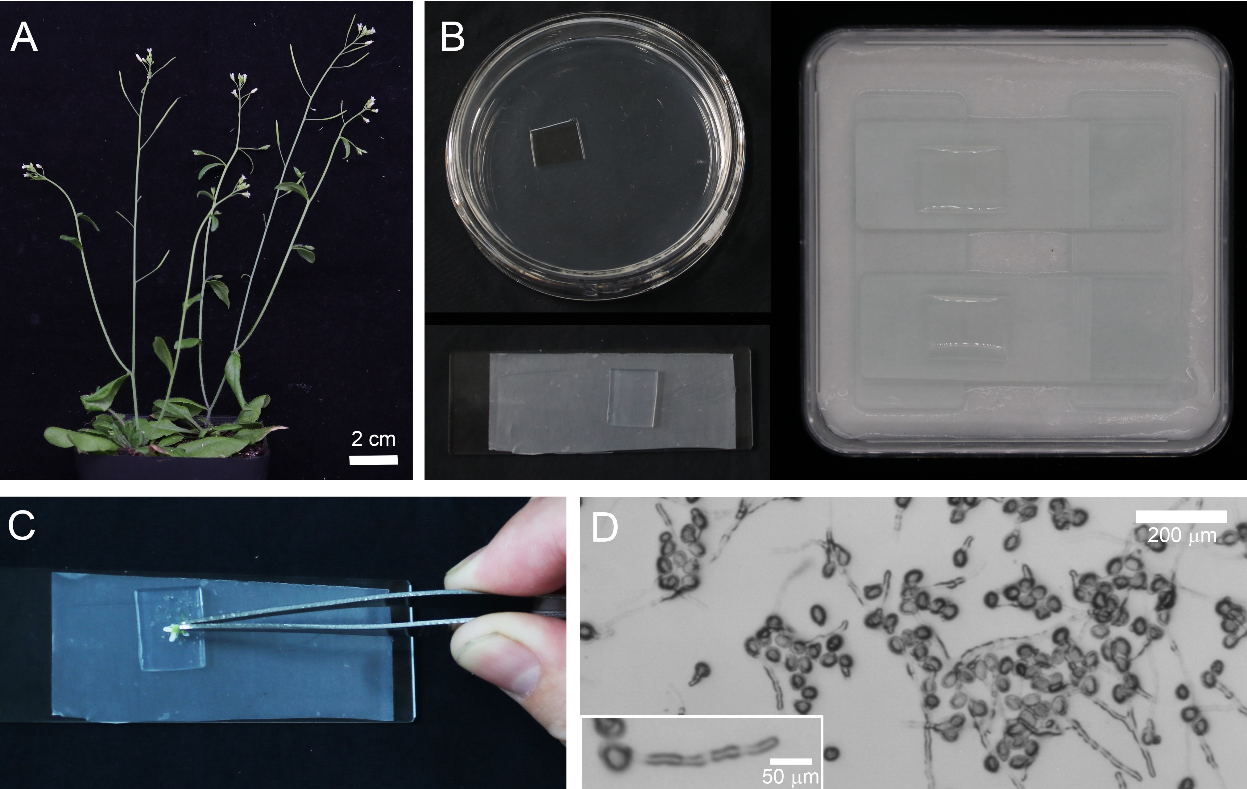
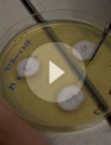
Figure 1. The Procedure for Germinating Arabidopsis Pollen on Solid Pollen Germination Medium. A. 6-8 weeks old Arabidopsis with flowers ready for the collection of pollen. Scale bar = 2 cm. B. Preparation of pollen germination medium and humid dish for pollen germination. C. Gently dip flowers to spread pollen on the surface of solid pollen germination medium. D.Images of pollen tubes after culture for 2 h at 28 °C. Scale bar = 200 μm. Inset is a pollen tube with the length reaches at ~180 μm. Scale bar = 50 μm. Pollen tubes are ready for subsequent fixation and staining with phalloidin. - Chemical reagents
- Calcium chloride (Merck/Sigma-Aldrich, catalog number: C7902)
- Calcium nitrate (Merck/Sigma-Aldrich, catalog number: C1396)
- Magnesium sulfate (Merck/Sigma-Aldrich, catalog number: M1880)
- Boric acid (Merck/Sigma-Aldrich, catalog number: B6768)
- Sucrose (Merck/Sigma-Aldrich, catalog number: V900116)
- Agarose (Merck/Sigma-Aldrich, catalog number: V900510)
- Potassium hydroxide (Merck/Sigma-Aldrich, catalog number: 484016)
- LatrunculinB (LatB) (Merck/Calbiochem, catalog number: 428020)
- Dimethyl sulfoxide (DMSO) (Merck/Sigma-Aldrich, catalog number: D2438)
- 3-Maleimidobenzoic acid N-hydroxysuccinimide ester (MBS) (Merck/Sigma-Aldrich, catalog number: M2786)
- Trizma (Merck/Calbiochem, catalog number: V900483)
- Sodium chloride (Merck/Sigma-Aldrich, catalog number: V900058)
- Nonidet P-40 (NP-40) (IGEPAL CA-630) (Merck/Sigma-Aldrich, catalog number: I-8896)
- Alexa Fluor 488 phalloidin (Thermo/Invitrogen, catalog number: A12379)
- Methanol (Merck/Sigma-Aldrich, catalog number: 494437)
- Milli-Q water (Merck)
- Pollen germination medium (see Recipes)
- Tris Buffered Saline (TBS) (see Recipes)
- Stock solution (see Recipes)
- Individual stocks with Milli-Q water (see Recipes)
Equipment
- Olympus FV1000S or an equivalent confocal microscope
- Olympus CX23 or an equivalent microscope
- Yiheng GHP-9050 or an equivalent constant temperature incubator
- 4 °C and -20 °C freezer
Software
- ImageJ (https://imagej.nih.gov/ij/ version 1.52n)
- R program (http://www.r-project.org)
Procedure
文章信息
版权信息
© 2020 The Authors; exclusive licensee Bio-protocol LLC.
如何引用
Qu, X., Wang, Q., Wang, H. and Huang, S. (2020). Visualization of Actin Organization and Quantification in Fixed Arabidopsis Pollen Grains and Tubes. Bio-protocol 10(1): e3509. DOI: 10.21769/BioProtoc.3509.
分类
植物科学 > 植物细胞生物学 > 细胞结构
植物科学 > 植物细胞生物学 > 细胞染色
您对这篇实验方法有问题吗?
在此处发布您的问题,我们将邀请本文作者来回答。同时,我们会将您的问题发布到Bio-protocol Exchange,以便寻求社区成员的帮助。