

Reconstituting Breast Tissue with Organotypic Three-dimensional Co-culture of Epithelial and Stromal Cells in Discontinuous Extracellular Matrices
通过不连续细胞外基质中上皮细胞和基质细胞三维共培养重建乳腺组织

发布: 2019年10月05日第9卷第19期 DOI: 10.21769/BioProtoc.3392 浏览次数: 5222
评审: Alak MannaWathsala WijayalathVarun Kesherwani
参见作者原研究论文
The authors used this protocol in:

Apr 2019


Abstract
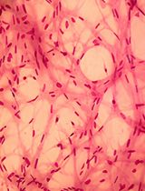
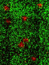
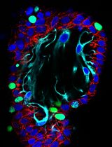
Co-culture systems utilizing reconstituted or synthetic extracellular matrix (ECM) and micropatterning techniques have enabled the reconstruction of surface epithelial tissues. This technique has been utilized in the regeneration, disease modeling and drug screening of the surface epithelia, such as the skin and esophagus. On the other hand, the reconstruction of glandular epithelia would require more intricate ECM organizations. Here we describe a protocol for a novel three-dimensional organotypic co-culture system for the reconstruction of mammary glands that utilizes the discontinuous ECM. In this technique, primary mammary fibroblasts first establish a layer of the connective tissue rich in collagen I. Then, mammary epithelial cells form acinar structures, the functional glandular units, within the laminin-rich basement membrane embedded in the connective tissue. This method allows for the regeneration of the in vivo-like architecture of mammary glands and could be utilized for monitoring the real-time response of mammary glands to drug treatment.
Keywords: ECM (ECM)Background
Three-dimensional (3D) co-culturing of cells in the extracellular matrix (ECM) substratum allows for the reconstruction of the in vivo-like tissue architecture and behavior, as well as the analysis of cellular interactions within the tissue microenvironment (Langhans, 2018). Furthermore, the advent of synthetic ECM materials and micropatterning techniques in the 2000s has enabled the regeneration of optimal ECM composition and spatial orientation for each tissue type in co-culture (Hotary et al., 2000; Gudjonsson et al., 2002; Carey et al., 2017). In these 3D co-culture systems, fibroblasts form a layer of the connective tissue while epithelial cells grow on top of the connective tissue or on the plastic surfaces exposed by micropatterning the ECM ( Stark et al., 2004; Kalabis et al., 2012; March et al., 2015). This method enables the reconstruction of 3D architecture of surface (stratified) epithelia, such as skin and esophagus. Such 3D co-culture technique has been utilized in generation of the transplantable skin organoids (Kim et al., 2018), disease modeling (e.g., psoriasis, esophagitis) and drug screening (Klicks et al., 2017; Whelan et al., 2018; Sarkiri et al., 2019). On the other hand, this technique is not suitable for the reconstruction of glandular epithelia that have more intricate ECM organizations. The glandular epithelium is enclosed by the basement membrane, which is, in turn, embedded in the connective tissue encompassing fibroblasts and other stromal cells.
Here we introduce a novel 3D organotypic ECM co-culture technique for reconstructing the 3D structure of the mammary gland. Mammary epithelial cells and mammary fibroblasts are individually cultured in the discrete, physiologically-relevant ECM. The discontinuous ECM is generated by micropatterning the fibroblast-containing collagen I-rich matrix and filling the grooves with the epithelial cell-containing laminin-rich basement membrane. This organotypic 3D ECM co-culture system allows for the growth of epithelial and stromal cells in the distinct locales, serving as a robust in vitro technique for modeling diseases and testing drug efficacy.
Materials and Reagents
- Materials
Flasks, plates and insert- T-75 flasks, vent (Corning, catalog number: 08-772-1F)
- Corning Cell Culture 12 well plates (Corning, catalog number: 07-200-82)
- Corning 12 mm Transwell with 3.0 μm Pore Polycarbonate Membrane Insert (Corning, catalog number: 07-200-157)
- Bio-wraps (Leica Biosystems, catalog number: 3801090)
- Tissue and Biopsy Cassettes (Fisher Scientific, catalog number: 15-200-403J)
Tools for Constructing a Custom Stamp- 5-minute epoxy (JB Weld, ClearWeld # 50112, Home Depot)
- Round wooden disks (25 mm in diameter, 3 mm in thickness, Hobby Lobby)
- Standard 2 mm drill bit
- Standard 5 mm drill bit
- Cell
- Primary human mammary epithelial cells (ScienCell, catalog number: 7610)
- MCF10A human mammary epithelial cells (Barbara Ann Karmanos Cancer Institute)
- HMT-3522-S1 human mammary epithelial cells (Mina Bissell Laboratory, Lawrence Berkeley National Laboratory, Berkeley, CA)
- Primary human mammary fibroblasts (ScienCell, catalog number: 7630)
- Media
For primary mammary epithelial cells- Mammary Epithelial Cell Medium (MEpiCM, ScienCell, catalog number: 7611) supplemented with 1% Mammary Epithelial Cell Growth Supplement (MEpiCGS, catalog number: 7652) and 1% penicillin/ streptomycin (Thermo Fisher, catalog number: MT30002CI)
For MCF10A human mammary epithelial cells- DMEM/F12 (Thermo Fisher, catalog number: 11320033) supplemented with the MCF10A additives (Recipe 1) (Debnath et al., 2003)
For HMT-3522-S1 human mammary epithelial cells- DMEM/F12 (Thermo Fisher, catalog number: 11320033) supplemented with the HMT-3522-S1 additives (Recipe 2) (Vidi et al., 2013)
For primary human mammary fibroblasts- Fibroblast Medium (FM, ScienCell, catalog number: 2301) supplemented with 1% Fibroblast Growth Supplement (FGS, catalog number: 2352) and 1% penicillin/streptomycin
- Reagents
Reagents for culturing- 0.05% trypsin/EDTA solution (Fisher Scientific, catalog number: 25-300-062)
- 0.25% trypsin/EDTA solution (Fisher Scientific, catalog number: 15-050-057)
- Soybean trypsin inhibitor (10 mg/ml, Thermo Fisher, catalog number: 17075-029)
- Dispase I (2mg [20U] Sigma-Aldrich, catalog number: D4818)
- 0.5 M EDTA, pH 8.0 (Thermo Fisher, catalog number: AM9260G)
- PBS (Thermo Fisher, catalog number: SH3037803)
- EGF (Sigma-Aldrich, catalog number: E-9644)
- Hydrocortisone (Sigma-Aldrich, catalog number: H-0888)
- Cholera toxin (Sigma-Aldrich, catalog number: C-8052)
- Horse serum (Thermo Fisher, catalog number: 16050122)
- Prolactin (Sigma-Aldrich, catalog number: L6520)
- β-estradiol (Sigma-Aldrich, catalog number: C-8052)
- Sodium selenite (Sigma-Aldrich, catalog number: 214485-5G)
- Transferrin (Sigma-Aldrich, catalog number: T2252)
- HMT-3522-S1 additives (see Recipes)
- MCF10A additives (see Recipes)
- Phosphate buffered saline (PBS) (see Recipes)
- Acellular layer matrix (see Recipes)
- Cellular layer matrix (see Recipes)
- Paraformaldehyde (see Recipes)
Reagents for ECM- Matrigel (~10 mg/ml, growth factor reduced, Corning, catalog number: 354230)
- Corning Collagen I, Rat Tail (3-4 mg/ml, Corning, catalog number: CB-40236)
- DMEM/F12 (10x), made from powder (Thermo Fisher, catalog number: 12500062)
- Sodium Bicarbonate 7.5% solution (50x, Thermo Fisher, catalog number: 25080094)
- FBS (JR Scientific, catalog number: 43603-500 [Research grade])
- L-Glutamine (200 mM [50x], Thermo Fisher catalog number: 25030081)
Reagents for Histology- Paraformaldehyde (Sigma-Aldrich, catalog number: P6148-500G)
Equipment
- Hand drill (De Walt, model: #dcd7916 20v 1/2" chuck, Home Depot)
- Stainless steel rods (Uxcell, 30 mm x 2.5 mm, Amazon.com)
- 4" cast iron drill press vice (Irwin, model: #226340, Home Depot)
- 37 °C water bath
- 37 °C humidified incubator with 5% CO2
- Cell culture benchtop centrifuge
- Hemocytometer
- Micropipettes (P20, P200 and P1000)
- Pipette aid
- Microscope with a color CCD camera
Procedure
文章信息
版权信息
© 2019 The Authors; exclusive licensee Bio-protocol LLC.
如何引用
Ren, G., Sharma, V., Letson, J., Walia, Y., Fernando, V. and Furuta, S. (2019). Reconstituting Breast Tissue with Organotypic Three-dimensional Co-culture of Epithelial and Stromal Cells in Discontinuous Extracellular Matrices. Bio-protocol 9(19): e3392. DOI: 10.21769/BioProtoc.3392.
分类
细胞生物学 > 细胞分离和培养 > 3D细胞培养
发育生物学 > 形态建成 > 器官形成
细胞生物学 > 细胞工程 > 组织工程
您对这篇实验方法有问题吗?
在此处发布您的问题,我们将邀请本文作者来回答。同时,我们会将您的问题发布到Bio-protocol Exchange,以便寻求社区成员的帮助。