

Explant Culture of the Embryonic Mouse Spinal Cord and Gene Transfer by ex vivo Electroporation
小鼠胚胎脊髓的组织块培养和体外电穿孔介导的基因转移


发布: 2019年09月20日第9卷第18期 DOI: 10.21769/BioProtoc.3373 浏览次数: 6970
评审: Giusy TornilloJackeline Moraes MalheirosAnonymous reviewer(s)
参见作者原研究论文
The authors used this protocol in:
Feb 2019


Abstract
Developing axons change responsiveness to guidance cues during the journey to synapse with target cells. Axon crossing at the ventral midline serves as a model for studying how axons accomplish such a switch in their response. Although primary neuron culture has been a versatile technique for elucidating various developmental mechanisms, many in vivo characteristics of neurons, such as long axon-extending abilities and axonal compartments, are not thoroughly preserved. In explant cultures, such properties of differentiated neurons and tissue architecture are maintained. To examine how the midline repellent Slit regulated the distribution of the Robo receptor in spinal cord commissural axons upon midline crossing and whether Robo trafficking machinery was a determinant of midline crossing, novel explant culture systems were developed. We have combined an “open-book” spinal cord explant method with that devised for flat-mount retinae. Here we present our protocol for explant culture of embryonic mouse spinal cords, which allows flexible manipulation of experimental conditions, immunostaining of extending axons and quantitative analysis of individual axons. In addition, we present a modified method that combines ex vivo electroporation and “closed-book” spinal cord explant culture. These culture systems provide new platforms for detailed analysis of axon guidance, by adapting gene knockdown, knockout and genome editing.
Keywords: Explant culture (组织块培养)Background
Growing axons sense numerous extracellular cues to reach their final target cells (Stoeckli, 2018). At intermediate targets on the way to the destination, axons timely switch on/off responses to guidance cues. Molecular mechanisms underlying this switch have been a hot topic in neurobiology (Guan and Rao, 2003; Sabatier et al., 2004; Dickson and Zou, 2010; Kolodkin and Tessier-Lavigne, 2011; Stoeckli, 2018; Yang et al., 2018; Ducuing et al., 2019).
The ventral midline of the central nervous system is an important intermediate target for different types of axons. Floor plate (FP) cells at the ventral midline secrete short- and long-range guidance cues, which act as attractants or repellents, and spatiotemporally organize the formation of neural circuits. Commissural axons project across the midline, connect the left and right sides of the nervous system and play roles in information transfer between both sides (Figures 1A and 1B). Commissural axons are initially guided to the midline by attractants derived from the FP and ventricular zones, such as netrin-1 and Sonic hedgehog (Dominici et al., 2017; Varadarajan et al., 2017; Moreno-Bravo et al., 2019; Wu et al., 2019). At the midline, commissural axons lose their responsiveness to the attractants and acquire responsiveness to repellents, such as Slit and semaphorins (Shirasaki et al., 1998; Zou et al., 2000). Thus, the midline is initially an attractive target for commissural axons, but becomes an unfavorable place upon their arrival, so that commissural axons exit the midline and never re-cross it. What mechanism ensures that commissural axons cross the midline only once? One key is the molecular basis by which commissural axons increase Slit sensitivity upon midline crossing.
Here we will summarize how spinal cord explant cultures have contributed to our understanding of axon guidance. The landmark research using explants showed that FP cells produce chemoattractant activity for commissural axons (Tessier-Lavigne et al., 1988). Two-dimensional co-culture assays using explants of commissural neurons and the FP revealed the presence of unknown midline repellent activity, which is unmasked by inhibiting either axonin-1/TAG-1 (a cell adhesion molecule [CAM] expressed in commissural axons) or NrCAM (expressed in FP cells) (Stoeckli et al., 1997). When Slit was identified as a ligand for Robo receptor and a long-awaited midline repellent (Brose et al., 1999; Kidd et al., 1999; Li et al., 1999), its repellent activity for vertebrate commissural axons was not clarified. But soon later, ingenious assay systems using FP-containing and FP-lacking spinal cord explants embedded in collagen gel revealed that Slit inhibits outgrowth of commissural axons that have crossed the midline (post-crossing axons), but not pre-crossing axons (Zou et al., 2000). This was the first demonstration that Slit is an evolutionally conserved midline repellent.
To examine how Slit sensitivity of commissural axons was regulated, we established a primary culture system (Kinoshita-Kawada et al., 2019; Yuasa-Kawada et al., 2009). Commissural neurons dissociated from dorsal spinal cords of mouse embryos at post-crossing stages (embryonic day [E]11.5-12.5) exhibit growth cone collapse and the increase in axonal Robo1 levels in response to Slit. In contrast, commissural neurons harvested from pre-crossing stage embryos show neither collapse responses to Slit nor the Slit-induced increase in axonal Robo1. Therefore, even without FP cells, dissociated commissural neurons recapitulate in vivo behaviors, with respect to the acquisition of Slit responsiveness. We then sought to develop an explant assay to examine Slit-induced Robo redistribution and growth cone responses in individual axons (or small fascicles) in a more physiological context, with subcellular resolution. Unfortunately, it was difficult to use so far developed explant assays for such purposes, because axons tended to form vigorous bundles in collagen gel.
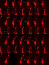
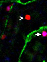
To study retinotectal topography, Drs. Friedrich Bonhoeffer, Uli Schwarz and colleagues developed retinal explant culture systems; a large mass of embryonic retinae were flat-mounted onto nitrocellulose (NC) filters and placed on the tectal cell membrane carpets, allowing robust extension of retinal axons (Halfter et al., 1981 and 1983; Bonhoeffer and Huf, 1982; Walter et al., 1987a and 1987b; Ichijo and Bonhoeffer, 1998; Yuasa-Kawada et al., 2003). By combining these protocols with spinal cord explant techniques (Zou et al., 2000), we prepared FP-lacking dorsal spinal cord [dSC (-FP)] and FP-containing hemisected spinal cord [SC (+FP)] explants (from cervical to lumbar levels) from E11.5 embryos and spread them on NC filters. Such explants were cultured on the Matrigel-coated substratum (Figures 1C-1G). Under these conditions, individual axons robustly extended from explants onto the two-dimensional surface, even without netrin-1, a stimulator of commissural axon growth. In both explant types, the extending axons were negative for TAG-1, a pre-crossing commissural axon marker but positive for L1, a post-crossing commissural axon marker (Dodd et al., 1988), maintaining the characteristics of post-crossing axons (Figure 3). These explant cultures allow simple immunostaining protocols and testing effects of Slit alone on Robo distribution (Kinoshita-Kawada et al., 2019).
Furthermore, to perform RNA interference (RNAi) against transducers of Slit-Robo signals, Arf6 GTPase and cytohesin Arf-guanine nucleotide exchange factors (Arf-GEFs), we modified an RNAi-based assay that combines ex utero electroporation with spinal cord explant culture (Wolf et al., 2008; Parra and Zou, 2010). Because the spinal cord, especially the FP region, becomes too fragile to prepare open-book explants after electroporation, we developed a new culture system of intact, “closed-book” spinal cords embedded in collagen gel (Kinoshita-Kawada et al., 2019) (Figures 2A-2D). The combination of these culture systems with RNAi-mediated knockdown or clustered regularly interspaced short palindromic repeats (CRISPR)-based genome editing, introduced by electroporation, virus or nanocomplex (e.g., Mikuni et al., 2016; Nishiyama et al., 2017; Park et al., 2019), will allow more flexibility to manipulate experimental conditions and to tag endogenous proteins. Such techniques will contribute to the elucidation of molecular mechanisms of axon guidance.
Materials and Reagents
- Animals
Pregnant ICR or C57BL/6J strain mice with embryonic day [E]~11.5 embryos
Note: We usually use 1-2 pregnant mice for each experiment. We assume an average of 6-12 embryos per ICR mouse and 4-8 embryos per C57BL/6J mouse. For timed-pregnancy mating, the day of vaginal plug was dated as E0.5, and embryos were staged according to EMAP (http://www.emouseatlas.org/emap/ema/home.php). In our hands, in terms of axon growth onto the Matrigel-coated substratum and the ease of handling, the best results have been obtained with embryos between E11.0 and E11.5, in order to prepare dSC (-FP) and SC (+FP) explants. At least, it is better to use embryos before E12.0 than later stages. For use of knockout mice, we prepare explants from littermate embryos, in parallel with genotyping. Image acquisition and analysis can be performed in a blinded manner. For ex vivo electroporation and closed-book explant culture, we recommend using ICR mouse embryos just before E11.5, with respect to the efficient introduction of axon-tracer plasmid/shRNA to commissural neurons prior to axon midline crossing and the maintenance of tissue integrity. - Surgical instruments and materials
- Scissors, large
- Scissors, medium
- Ophthalmic scissors (option) (Natsume Seisakusho, catalog number: MB-50-7)
- Forceps (7) (Dumont, No. 5 Inox)
Note: We use relatively blunt forceps (a total of 5; 1 for handling coverslips, 2 for mouse surgery and 2 for spinal cord dissection) and sharpened forceps for fine dissection (2). - Straight metal spatula, with polished edges
- Disposable scalpel (Feather, No. 11)
- Sharpened tungsten needles, bent at a right angle (tungsten wire, diameter: 0.20 mm; Nilaco, catalog number: W-461267) and needle holders (Fine Science Tools, catalog number: 26016-12) (see Figure 1H).
Note: At the tip, the diameter should be approximately 20 μm. The needles can be sharpened in a 1 N NaOH bath by putting the negative electrode of a power supply (low voltage should be set) into the bath and the positive electrode (alligator clip) onto the end of needle holder. Dip in/out the needle tip to make a pointed, tapered tip. See the Recipe “Sharpened tungsten needles” in Cold Spring Harb Protoc (doi:10.1101/pdb.rec069468). - Disposable transfer pipettes, 3.5 ml (Sarstedt, catalog number: 86.1171.001) or equivalent (approximately 15 cm in length, with an inner diameter of 2 mm)
Note: When handling whole spinal cords, use cut pipettes, which have opening of approximately 4-5 mm. When handling excised, tiny spinal cord explants, we usually use un-cut pipettes. - Acid-washed, sterilized, round coverslips (18 mm in diameter; Matsunami, thickness: No. 1; for preparing the coverslips, see Recipe 1)
- Black nitrocellulose (NC) membrane filter (gridded on one surface, single-packed and sterile) (Sartorius, Cellulose nitrate filter, catalog number: 13006--47----CAN)
- Glass slides (Matsunami, Superfrost, catalog number: S2443)
- 70% Ethanol
- Culture products
- 35-mm sterile tissue culture dishes (Corning, Falcon, catalog number: 353001)
- 60-mm sterile tissue culture dishes (Corning, Falcon, catalog number: 353004)
- 100-mm sterile tissue culture dishes (Corning, Falcon, catalog number: 353003)
- 15-ml conical centrifuge tubes (Thermo Fisher Scientific, catalog number: 14-959-70C)
- 50-ml conical centrifuge tubes (Eppendorf, catalog number: 0030122178)
- 1.5-ml safe-lock microcentrifuge tubes (Eppendorf, catalog number: 0030120086)
- Metal weights (stainless steel; made by a local machine shop)
Note: Before use, wash the weights at least three times with distilled water (washing with sonication for 5-10 min is preferable) and with 100% ethanol overnight, air-dry and sterilize them by baking at 180 °C for 3 h or by UV irradiation (when hurrying). It is convenient to have many metal weights of various sizes, although we usually use the 3 x 3 x 14 mm-size weights (see Figure 1M). After use, wash them three times with distilled water (and perform sonication) and with 100% ethanol overnight. Follow the above sterilization procedure for the next round of use. Avoid using detergent for washing and do not autoclave them. - Matrigel matrix (Corning, catalog number: 354234)
Note: Thaw a bottle of Matrigel overnight on ice. Make aliquots (0.2 ml per 1.5 ml tube) and store at -80 °C. - Concanavalin A (ConA; Type IV, lyophilized powder; Sigma-Aldrich, catalog number: C2010-25MG)
- ConA stock solution (see Recipe 2)
- Culture media
- Hanks’ balanced salt solution, no calcium, no magnesium (HCMF; Thermo Fisher Scientific, catalog number: 14170112)
- Dulbecco’s modified Eagle’s medium (DMEM), high glucose (Thermo Fisher Scientific, catalog number: 11965092)
- L-Glutamine, 200 mM solution (Thermo Fisher Scientific, catalog number: 25030081)
- Fetal bovine serum (FBS) (Thermo Fisher Scientific, Gibco, catalog number: 10270106 or Hyclone, catalog number: SH30910.03)
Note: Heat-inactivation of FBS at 56 °C for 30 min is not essential in this culture protocol. - Neurobasal medium (Thermo Fisher Scientific, catalog number: 21103049)
- B-27 supplement (50x), serum-free (Thermo Fisher Scientific, catalog number: 17504044)
- Opti-MEM I reduced serum medium (Thermo Fisher Scientific, catalog number: 31985062)
- Penicillin-streptomycin, liquid (designated Pen/Strep; used as 100x solution; 10,000 units/ml each) (Thermo Fisher Scientific, catalog number: 15140122)
- HEPES (1 M) (Thermo Fisher Scientific, catalog number: 15630080)
- Purified human recombinant Slit2 (option: used in our experiment; store aliquots at -80 °C)
Note: Recombinant Slit2 proteins were purified from conditioned media from HEK293 cells stably expressing human Slit2-myc (Li et al., 1999) by ion-exchange chromatography using SP Sepharose Fast Flow (GE Healthcare) (Guan et al., 2007). Control purified preparations, made from parental HEK293 cells by employing the same procedure as for Slit purification, were used for explant assays in parallel. Our detailed protocol for Slit purification is available upon request to the authors. - Medium 1 (see Recipe 3)
- Medium 2 (see Recipe 4)
- Medium 3 (see Recipe 5)
- Materials for ex vivo electroporation and closed-book spinal cord explant culture
- Glass capillaries (Narishige, catalog number: GD-1)
Note: To prepare injection pipettes, pull glass capillaries using a vertical puller and break the capillary tip with fine forceps; if possible, cut it diagonally (tip diameter: approximately 30 μm). - Vertical puller (Narishige, current model: PC-100)
- Mouthpiece and pipette joints (for injection before electroporation, we use an apparatus available from Suisaku, Japan (model: slim tube), to which an appropriate length of silicon tube can be attached)
- DPBS, no calcium, no magnesium (Thermo Fisher Scientific, catalog number: 14190144)
- Leibovitz's L-15 (Thermo Fisher Scientific, catalog number: 11415064)
- Horse serum, heat-inactivated (Thermo Fisher Scientific, catalog number: 26050070)
- Axon tracer (EGFP/vYFP plasmid)
Note: We have compared among pCAG-EGFP (available from Nepa Gene), pCAG-vYFP (Venus), pCAG-GAP43-EGFP and pCAG-GAP43-vYFP for axon labeling. In our study, pCAG-vYFP showed the best performance in mouse spinal cords. We usually obtain midi-prep grade DNAs by using NucleoBond Xtra Midi EF kit (Macherey-Nagel). We then clean up the DNAs by conventional phenol-chloroform extraction and ethanol precipitation, and obtain a high-purity DNA stock by dissolving in sterile, endotoxin-free TE buffer at a concentration of 8-10 μg/μl. Store at -30 °C (avoid thawing/refreezing cycles of the stock too many times). - Appropriate short-hairpin RNAs (shRNAs; in this paper, we used shRNA against Arf6 [shArf6] cloned in pRS shRNA vector; Origene) and control shRNA (shControl) for RNAi-mediated knockdown (the DNAs are purified as described above and dissolved at an appropriate concentration; see Kinoshita-Kawada et al., 2019)
- 0.5-1% Fast Green (10x) (Sigma-Aldrich, catalog number: F7252)
Note: After dissolving in DPBS, sterilize by filtration (0.22 μm). Make aliquots and store them at -30 °C. - Native collagen acidic solution I-AC (3 mg/ml, pH3.0) (Koken, catalog number: IAC-30)
- DMEM/F-12, powder (Thermo Fisher Scientific, catalog number: 12500062)
- 5x concentrated DMEM/F-12 medium (Recipe 6)
- Reconstitution buffer (used for gelatinization of collagen; see Recipe 7)
- Medium 4 (see Recipe 8)
- Glass capillaries (Narishige, catalog number: GD-1)
- Fixation
- PBS (Phosphate-buffered saline) tablets (Takara, catalog number: T900)
Note: Sterility is not required, although the above DPBS can also be used. - PBS + 0.025% NaN3
- Paraformaldehyde (PFA) (Fuji film/Wako, catalog number: 162-16065)
- 4% PFA in PBS supplemented with 10% sucrose (~0.3 M sucrose; Walter et al., 1987a; for fixing cultured explants without electroporation)
Note: Prepare a stock in a concentration of 4% PFA in PBS, make aliquots (approximately 30 ml per 50 ml tube) and store at -30 °C. For use in fixation, thaw 4% PFA in PBS and add sucrose to approximately 10 %. - 4% PFA in PBS (for fixing cultured explants of electroporated spinal cords)
- PBS (Phosphate-buffered saline) tablets (Takara, catalog number: T900)
- Immunostaining and observation
- 24 x 60 mm No. 1 Micro cover glass (Matsunami, catalog number: C024601)
Note: This coverslip is used for mounting spinal cord explants that are electroporated and cultured. - Sylgard-coated dissection dishes (for preparation, see page 275 of Banker and Goslin, 1998)
- 0.2% Triton X-100 in PBS
Note: We freshly dilute Triton X-100 (Sigma-Aldrich, catalog number: T8787) on the day of immunostaining. - Bovine serum albumin (BSA) (Sigma-Aldrich, catalog number: A2153)
- Goat serum (Sigma-Aldrich, catalog number: G6767)
- Skim milk (BD/Difco, catalog number: 232100)
- Rabbit primary antibodies of interest (in our study, we use anti-Robo antibodies)
- Anti-L1CAM (mouse monoclonal IgG1; clone 2C2; Abcam, catalog number: ab24345)
- Anti-TAG-1 (mouse monoclonal IgM; clone 4D7; Developmental Studies Hybridoma Bank)
- Alexa Fluor 647 mouse anti-β-tubulin, class III (clone TuJ1; BD, catalog number: 560394)
- Goat anti-mouse IgG (H+L) highly cross-adsorbed secondary antibody, Alexa Fluor 555 (Thermo Fisher Scientific, catalog number: A32727)
- Goat anti-mouse IgM (heavy chain) cross-adsorbed secondary antibody, Alexa Fluor 555 (Thermo Fisher Scientific, catalog number: A21426)
- Goat anti-rabbit IgG (H+L) highly cross-adsorbed secondary antibody, Alexa Fluor 488 (Thermo Fisher Scientific, catalog number: A11034)
- PermaFluor aqueous mounting medium (Thermo Fisher Scientific, catalog number: TA-030-FM)
Equipment
- CO2 tank
- CO2 incubator (ASTEC, water-jacket type)
- CO2 single-stage flowmeter regulator
- Dissecting microscope (Carl Zeiss, model: Stemi 2000-C; current models: Stemi 305/508)
- Cold light source (FOSTEC, model: DCRII)
- Phase contrast microscope (Olympus, model: CKX31; current model: CKX53)
- Millipore Milli-Q ultrapure water system
- Laminar flow hood
- 4 °C refrigerator
- -30 °C freezer
- -80 °C freezer
- Electroporator (Nepa Gene, model: NEPA21)
Note: NEPA21 is a versatile electroporator, which is highly programmable and reproducible with many variables controlled and measurable. - Forceps-type electrodes (to electroporate mouse spinal cords, we preferably use CUY665P9-6-2-5, with the tip diameter of 2.5 mm, Nepa Gene)
Note: The shape of electrode tips remarkably affects the efficiency and pattern of gene transfer into the spinal cord. - Confocal laser-scanning microscope (Carl Zeiss, model: LSM780 or LSM880) or all-in-one fluorescence microscope (Keyence, model: BZ-X700)
Note: If possible, use an appropriate confocal microscope with image-tiling functions (see Figure 3A). Although laborious, montage images can also be assembled manually. In our initial studies, we used a wide-field BX61I microscope (Olympus) equipped with a CoolSNAP HQ or ES CCD camera (Roper Industries). We manually assembled images that were processed with AutoQuant X3 deconvolution software (Media Cybermetrics; see Figure 3C).
Software
- MetaMorph (version 7.7 and 7.10; Molecular Devices)
- Photoshop CS5 Extended (Adobe)
- Illustrator CS5 (Adobe)
- Excel (Microsoft)
- Prism 6 (GraphPad)
Procedure
文章信息
版权信息
© 2019 The Authors; exclusive licensee Bio-protocol LLC.
如何引用
Kinoshita-Kawada, M., Hasegawa, H., Hongu, T., Yanagi, S., Kanaho, Y., Masai, I., Mishima, T., Chen, X., Tsuboi, Y., Rao, Y., Yuasa-Kawada, J. and Wu, J. Y. (2019). Explant Culture of the Embryonic Mouse Spinal Cord and Gene Transfer by ex vivo Electroporation. Bio-protocol 9(18): e3373. DOI: 10.21769/BioProtoc.3373.
分类
神经科学 > 发育 > 外植体培养
神经科学 > 发育 > 电穿孔
细胞生物学 > 细胞运动 > 细胞迁移
您对这篇实验方法有问题吗?
在此处发布您的问题,我们将邀请本文作者来回答。同时,我们会将您的问题发布到Bio-protocol Exchange,以便寻求社区成员的帮助。