- EN - English
- CN - 中文
Immunoelectron Microscopy in Chlamydomonas Cells
免疫电镜术检测衣藻属细胞
发布: 2013年03月05日第3卷第5期 DOI: 10.21769/BioProtoc.335 浏览次数: 12898

相关实验方案

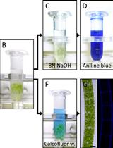
采用苯胺蓝和Calcofluor White对绿藻类双星藻属和克里藻属的胼胝质和纤维素进行染色
Klaus Herburger and Andreas Holzinger
2016年10月20日 14721 阅读
Abstract
The method of immunoelectron microscopy is intended for localization of proteins inside the cells of Chlamydomonas reinhardtii or other microalgae and cyanobacteria. This protocol was used to study localization of carbonic anhydrase Cah3 with antibodies raised in rabbit, though it can be used to localize any other abundant protein. Primary rabbit antibodies are recommended because they react quickly and specifically with proteins of C. reinhardtii. If primary antibodies other than rabbit are used, the blocking procedure and time of incubation with primary and secondary antibodies should be adjusted.
Keywords: Ultrastructure (超微结构)Materials and Reagents
- Culture of C. reinhardtii 137mt+ (WT) (we used the strain IPPAS D-298 from the Collection of microalgae of the Institute of Plant Physiology RAS, Moscow)
- Primary antibodies raised in rabbits against the protein of interest (we used antibodies raised against the recombinant Cah-3 protein (α-CA) of C. reinhardtii, Agrisera, Vannas, Sweden)
- Secondary antibodies: Anti-Rabbit IgG conjugated with 10 nm colloidal gold particles (Sigma-Aldrich, catalog number: G7402 )
- Paraformaldehyde (Sigma-Aldrich, catalog number: P6148 )
- LR White embedding kit (Sigma-Aldrich, catalog number: 62662-1EA-F )
- Gelatin capsules (SPI supplies, catalog number: 02308-SS )
- Formvar and carbon-coated nickel grids (SPI supplies, catalog number: 3430N-CF )
- Bovine serum albumin (BSA) (Sigma-Aldrich, catalog number: A7906 )
- Goat serum (Sigma-Aldrich, catalog number: G9023 )
- Rabbit serum (Sigma-Aldrich, catalog number: R4505 )
- Paraformaldehyde (Sigma-Aldrich, catalog number: P6148 )
- Ethanol
Optional - Uranyl acetate (e.g. SPI supplies, catalog number: 02624-AB )
- Lead citrate (e.g. Sigma-Aldrich, catalog number: 15326 )
- NaOH
- Phosphate buffer saline (PBS) (pH 7.4) (see Recipes)
- Fixation solution (4% paraformaldehyde) (see Recipes)
- Tris-buffer saline (TBS) (pH 7.4) (see Recipes)
- 1% BSA-TBS (see Recipes)
- Uranyl acetate (see Recipes)
- Lead citrate (see Recipes)
Equipment
- 96 well immunology plate (e.g. Greiner Bio-One, catalog number: 650001 )
- Pasteur pipettes with thin tips (ROTHE 306.1)
- Thermostat unit with, at least, 37-55 °C temperature range (e.g. BINDER B28)
- Ultramicrotome (e.g. Reichert, OMU-3, Austria)
- Grid storage box (Sigma-Aldrich, catalog number: G6276 )
- Transmission electron microscope (e.g. Libra-120, Carl Zeiss, Germany)
- Plastic Petri dish or Teflon plate with hydrophobic surface
- Wet chamber
Procedure
文章信息
版权信息
© 2013 The Authors; exclusive licensee Bio-protocol LLC.
如何引用
Sinetova, M. A. and Markelova, A. G. (2013). Immunoelectron Microscopy in Chlamydomonas Cells. Bio-protocol 3(5): e335. DOI: 10.21769/BioProtoc.335.
分类
植物科学 > 藻类学 > 蛋白质
植物科学 > 藻类学 > 细胞分析
细胞生物学 > 细胞染色 > 蛋白质
您对这篇实验方法有问题吗?
在此处发布您的问题,我们将邀请本文作者来回答。同时,我们会将您的问题发布到Bio-protocol Exchange,以便寻求社区成员的帮助。
Share
Bluesky
X
Copy link


