

Photodynamic Therapy in a 3D Model of Ovarian Cancer
卵巢癌3D模型中的光动力学治疗

发布: 2019年08月05日第9卷第15期 DOI: 10.21769/BioProtoc.3314 浏览次数: 5625
评审: Khyati Hitesh ShahAnne-Marie Caroline OverstreetAnonymous reviewer(s)
参见作者原研究论文
The authors used this protocol in:

Jan 2019
Advertisement


Abstract
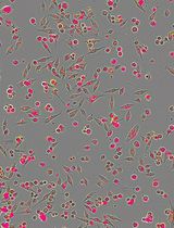
Photodynamic therapy (PDT), is a clinically-approved light-based anti-cancer treatment modality in which a photoactivatable photosensitizer is irradiated with an appropriate wavelength of light to generate cytotoxic molecules to kill cancer cells. In this article, we describe an in vitro PDT protocol using a 3-dimensional (3D) model of ovarian cancer that was established on beds of Matrigel. PDT was performed using a liposomal formulation of verteporfin photosensitizer (Visudyne®). The cancer cells were genetically-labeled with the fluorescent protein mCherry to facilitate the evaluation of the treatment response. This protocol is advantageous because the mCherry fluorescence is an indicator of cell viability, eliminating the need for external dyes, which often exhibit limited penetration and diffusion into 3D organoids. Additionally, Visudyne PDT achieves significant tumor-killing efficacy in a 3D model for ovarian cancer.
Keywords: Photodynamic therapy (光动力疗法)Background
Photodynamic therapy (PDT) is an FDA-approved light-based treatment modality that utilizes a photoactivatable molecule, termed a photosensitizer (PS). Upon irradiation with an appropriate wavelength of light, the PS generates reactive molecular species (RMS) within the cytosol. These RMS, often reactive oxygen species, induce photodamage and are toxic to the cell (Celli et al., 2010; Obaid et al., 2016). Certain formulations of PS preferentially accumulate in the malignant tissue and thus PDT often achieves dual selectivity as a result of confined PS accumulation in the tumor and its spatiotemporal control over the light irradiation. PDT is clinically approved for several indications, including esophageal cancer, lung cancer, age-related macular degeneration, and is being investigated for many more cancerous and noncancerous indications (Celli et al., 2010).
Ovarian cancer is a deadly gynecologic malignancy often associated with treatment-resistant metastases and poor outcomes in patients. While surgery and chemotherapy are the current standard of care for ovarian cancer, the efficacy of PDT to manage ovarian cancer is currently being investigated in pre-clinical studies by many research groups (Goff et al., 1996; Rizvi et al., 2010).
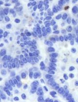
Like many other malignancies, ovarian cancer metastases display complex morphological and biochemical characteristics that are lost in traditional monolayer (2D) cell culture. 3D tumor model is an emerging technique for evaluating tumor properties and treatment response in vitro. 3D cultures of tumor cells create a more physiologically relevant tumor microenvironment than traditional 2D cell culture because they retain the biological and structural features of tumors that are found in vivo (Rizvi et al., 2016). Cells in monolayer experience contact inhibition and a homogeneous oxygen and nutrient environment, while 3D cultures display heterogeneity within and across spheroids and restore extracellular matrix-related cues that are important in treatment response. Like in vivo tumors, 3D cultured tumors proliferate into heterogeneous clusters with varying levels of oxygen and nutrients, making them useful tools for studying PS uptake and PDT efficacy within oxygen gradients. Increased PDT resistance has been found in 3D tumor models compared with 2D, providing a platform for testing alternative treatment regimens and combination therapies to overcome this resistance. The complex factors that contribute to treatment failure in patients necessitate an in vitro model that mimics the structural, biological, and extracellular cues that play a major role in treatment outcomes (Rizvi et al., 2016).
Here, we describe an in vitro PDT protocol using a 3D model of ovarian cancer. 3D nodules of human ovarian cancer cells (NIH: Ovcar5) that stably express a fluorescent reporter were grown on beds of Growth Factor-Reduced Matrigel®. PDT was performed using FDA-approved PS verteporfin (Visudyne®). The treatment response was evaluated by imaging of the fluorescent 3D nodules. The protocol described here has been adapted from a previously published article (Rizvi et al., 2019).
Materials and Reagents
- Pipette tips (200 and 1,000 μl, Fisher Scientific, catalog numbers: 02-707-407 and 02-707-413, respectively)
- LASER-safety glasses (Thorlabs, catalog number: LG3)
- Aspirating pipettes (Celltreat, catalog number: 229265)
- 5 and 10 ml serological pipettes (Corning, catalog numbers: 357543 and 357551, respectively)
- 15 ml centrifuge tubes (Corning, catalog number: 352095)
- 50 ml centrifuge tubes (Corning, catalog number: 430829)
- 24-well glass-bottom black-wall plate (Greiner Sensoplates, catalog number: 662892)
- T75 flask (Corning, catalog number: 353136)
- Aluminum foil (Fisher Scientific, 01-213-105)
- mCherry-expressing NIH: OVCAR5 cell line (Rizvi et al., 2019) (“mCherry-Ovcar5” thereafter)
- Visudyne® (Verteporfin for injection by Bausch & Lomb)
- Dulbecco’s Phosphate Buffered Saline (DPBS) without Ca2+ & Mg2+ (Corning, Cellgro, catalog number: 21-031-CV)
- Heat inactivated fetal bovine serum (FBS) (Gibco, catalog number: 26140079)
- 100x Penicillin-streptomycin solution (Corning, catalog number: 30001Cl)
- Trypsin-EDTA solution (Corning Cellgro, catalog number: 25-052-Cl)
- Corning Growth factor-reduced (GFR) Matrigel® Matrix (“Matrigel” thereafter) (Corning, catalog number: 354230)
- Matrigel matrix using Cell Recovery Solution (Corning, catalog number: 354253)
- RPMI 1640 medium (Corning, Cellgro, catalog number: 10-040-CV)
- DMSO (Sigma, catalog number: D2438)
- RPMI Growth Medium (see Recipes)
- 4% Matrigel (see Recipes)
- Visudyne® working solution (see Recipes)
Equipment
- Pipettes (P1000 and P200, Gilson, catalog numbers: F123602 and F123601, respectively)
- Biosafety cabinet for cell culture (Baker Company, catalog number: SG-200)
- Pipette-aid (Biotang, catalog number: 710932)
- Analytical balance (Sartorius, catalog number: ENTRIS2241SUS)
- Inverted light microscope (VWR, catalog number: 89404-462)
- 5x air objective (NA 0.16) lens (PerkinElmer, catalog number: HH14000402)
- Freezer (-20 °C) (Thermo Scientific, model: 20LFEETSA)
- LASER Source (690 nm) (Intense Ltd., model: Intense series 7400)
- Laser power meter (Ophir Vega, catalog number: 7Z01560)
- Laser shutter controller (INLINE-TTL Electronic Shutter)
- Centrifuge machine (Beckman, model: Allegra 6R)
- High-Content Imaging Instrument (Operetta CLS, PerkinElmer)
- Humidified cell incubator (Thermo Scientific, model: FormaTM 310)
- Cell counter (Beckman Coulter, model: Z1 Particle Counter)
Software
- Harmony 4.6 (PerkinElmer)
- GraphPad Prism 8
- (Optional) ImageJ or qVista software
Procedure
文章信息
版权信息
© 2019 The Authors; exclusive licensee Bio-protocol LLC.
如何引用
Nath, S. and Moore, K. (2019). Photodynamic Therapy in a 3D Model of Ovarian Cancer. Bio-protocol 9(15): e3314. DOI: 10.21769/BioProtoc.3314.
分类
癌症生物学 > 通用技术 > 癌症治疗
细胞生物学 > 细胞活力 > 细胞死亡
您对这篇实验方法有问题吗?
在此处发布您的问题,我们将邀请本文作者来回答。同时,我们会将您的问题发布到Bio-protocol Exchange,以便寻求社区成员的帮助。