

Imaging Gene Expression Dynamics in Pseudomonas fluorescens In5 during Interactions with the Fungus Fusarium graminearum PH-1
荧光假单胞菌In5与小麦赤霉菌PH-1互作过程中的基因表达动态成像


发布: 2019年06月20日第9卷第12期 DOI: 10.21769/BioProtoc.3264 浏览次数: 5357
评审: Juan Facundo Rodriguez AyalaFernando A Gonzales-ZubiateJose Antonio Reyes-Darias
参见作者原研究论文
The authors used this protocol in:

Mar 2017


Abstract
Genomics, transcriptomics and metabolomics are powerful technologies for studying microbial interactions. The main drawback of these methods is the requirement for destructive sampling. We have established an alternative but complementary technique based on a microplate system combined with promoter fusions for visualizing gene expression in space and time. Here we provide a protocol for measuring spatial and temporal gene expression of a bacterial reporter strain interacting with a fungus on a solid surface.
Keywords: Microbial interactions (微生物相互作用)Background
Microbial interactions underpin many important biotechnological applications spanning medicine, food development and processing, bioremediation and biocontrol. In order to study microbial interactions, a broad range of technologies exist including genomics, transcriptomics and mass spectrometry imaging to study gene expression and metabolites exchanged during interactions. While these methods advance our understanding of microbial interactions, they also have limitations namely the requirement for destructive sampling. We have developed a microplate reader-based system for visualizing gene expression dynamics in living bacterial cells in response to a fungus in space and real-time. Pseudomonas fluorescens In5 is a Gram-negative soil bacterium and potent producer of secondary metabolites with antifungal activity (Michelsen et al., 2015; Hennessy et al., 2015). We previously identified the LuxR-type regulator NunF as a key regulator for synthesis of the antifungal compounds nunamycin and nunapeptin (Hennessy et al., 2017a). In this protocol, we detail how in vivo monitoring of target gene expression in living bacterial cells interacting with living fungal cells can be performed using a microplate-reader based technique (Hennessy et al., 2017b). P. fluorescens In5 expressing the red fluorescent protein mCherry fused to the promoter region of a regulator gene nunF indicating activation of an antifungal secondary metabolite gene cluster was used as a reporter system. Time-lapse image recordings of the reporter red signal and a green signal from fluorescent metabolites naturally produced by the bacterium combined with microbial growth measurements showed that nunF-regulated gene transcription is switched on when the bacterium enters the deceleration growth phase and upon physical encounter with fungal hyphae. The established non-destructive method has many advantages notably the ability to provide detailed space and time information on the transcription of target genes in living organisms. Importantly, the technique is a fast and simple alternative and complementary tool to the many technologies already used for studying microbial interactions.
Here we present a detailed protocol for imaging microbial interactions. In this example, we describe the construction of a reporter strain of the antimicrobial isolate Pseudomonas fluorescens In5 coupled with imaging analysis of gene expression of the reporter strain during an interaction with the fungus Fusarium graminearum PH-1 on an agar surface.
Materials and Reagents
- Eclipse pipette refilling system (Labcon, catalog numbers: 1045-260-000, 1093-260-000, 1036-260-000)
- NuncTM OmniTrayTM Single-Well Plate (Fischer Scientific, catalog number: 140156)
- Gene Pulser® Cuvettes 0.2 cm gap (Bio-Rad, catalog number: 1652086)
- 2 ml Microcentrifuge tubes (Fischer Scientific, catalog number: 21-402-905)
- NEB® 5-alpha Competent E. coli (Subcloning Efficiency) (NEB, catalog number: C2988J)
- Restriction enzymes: BamHI (NEB, catalog number: R3136S ), EcoR1 (NEB, catalog number: R0101S )
- Gentra Puregene Yeast/Bact. kit (QIAGEN, catalog number: 158567)
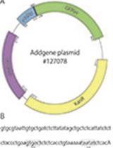
- Reporter plasmid pSEVA237R expressing the red fluorescent protein mCherry (Hennessy et al., 2018)
- Phusion high-fidelity polymerase (Fisher Scientific, catalog number: F530S)
- Wizard® SV Gel and PCR Clean-Up System (Promega, catalog number: A9281)
- Plasmid Mini Kit QIAprep Spin Miniprep Kit (QIAGEN, catalog number: 27104)
- Gibson Assembly® Cloning Kit (NEB, catalog number: E5510S)
- BactoTM agar (Difco, catalog number: 214050)
- Luria-Bertani broth (LB) (Difco, catalog number: 244610)
- LB agar (LBA) (Difco, catalog number: 244520)
- SOC outgrowth medium (NEB, catalog number: B9020S)
- Defined Fusarium Medium (DFM) (Hennessy et al., 2017b)
- Kanamycin sulfate monohydrate (Duchefa Biochemie, catalog number: K0126)
- Sucrose (Sigma-Aldrich, catalog number: S0389)
- Bacto-tryptone (VWR Chemicals, catalog number: 84610)
- Yeast extract (VWR Chemicals, catalog number: 84601)
- NaCl (Millipore, catalog number: 1.06404.1000)
- Glucose (Sigma-Aldrich, catalog number: 49159)
- Asparagine (Difco, catalog number: 214410)
- MgSO4 (VWR Chemicals, catalog number: 25164.265)
- KH2PO4 (Sigma-Aldrich, catalog number: NIST200B)
- KCl (Sigma-Aldrich, catalog number: P9333)
- Na2B4O7·10H2O (Sigma-Aldrich, catalog number: S9640)
- CuSO4·5H2O (Sigma-Aldrich, catalog number: C8027)
- FeSO4·7H2O (Sigma-Aldrich, catalog number: 1270355)
- MnSO4·H2O (Sigma-Aldrich, catalog number: M7634)
- NaMoO2·2H2O (Sigma-Aldrich, catalog number: 331058)
- ZnSO4·7H2O (Sigma-Aldrich, catalog number: Z0251)
- 300 mM sucrose (see Recipes)
- LB medium (see Recipes)
- DFM agar (see Recipes)
- 1,000x Trace solution (see Recipes)
Equipment
- Pipettes (VWR, catalog numbers: 613-2696E, 613-2702E, 613-2704E)
- Microplate reader (BMG LABTECH, catalog number: FLUO-star Omega)
- Eppendorf® Mastercycler® (Sigma-Aldrich, catalog number: EP6313000018-1EA)
- Gene Pulser® II (Bio-Rad, catalog number: 165-2105)
- Sterile inoculation loops (10 μl) (VWR, catalog number: 12000-812)
- Cork borer (6 mm Ø) for making fungal plugs
- Water bath or a heat block
Software
- BMG Omega Mars Software (Omega V5.11; BMG LABTECH)
- ImageJ analysis software (https://imagej.nih.gov/ij/)
- "Text image to stack” macro (ImageJ, http://rsbweb.nih.gov/ij/macros/ImportTextImageSequence.txt)
- Bulk Rename Utility, (https://www.bulkrenameutility.co.uk)
- Batch converter software converting BMG Omega Mars exported well scan text files to image text files for a rectangle of adjacent wells (MofM.exe)
Procedure
文章信息
版权信息
© 2019 The Authors; exclusive licensee Bio-protocol LLC.
如何引用
Hennessy, R. C., Stougaard, P. and Olsson, S. (2019). Imaging Gene Expression Dynamics in Pseudomonas fluorescens In5 during Interactions with the Fungus Fusarium graminearum PH-1. Bio-protocol 9(12): e3264. DOI: 10.21769/BioProtoc.3264.
分类
微生物学 > 微生物遗传学 > 基因表达
分子生物学 > 蛋白质 > 表达
您对这篇实验方法有问题吗?
在此处发布您的问题,我们将邀请本文作者来回答。同时,我们会将您的问题发布到Bio-protocol Exchange,以便寻求社区成员的帮助。