

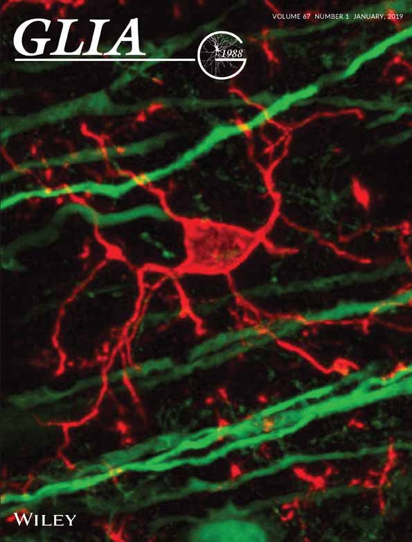
Time-lapse Whole-field Fluorescence Imaging of Microglia Processes Motility in Acute Mouse Hippocampal Slices and Analysis
急性海马切片中小胶质细胞运动长时间全视野荧光成像及分析


(*contributed equally to this work) 发布: 2019年04月20日第9卷第8期 DOI: 10.21769/BioProtoc.3220 浏览次数: 6661
评审: Xi FengXiaoliang ZhaoAnonymous reviewer(s)
参见作者原研究论文
The authors used this protocol in:
Jan 2019
Advertisement


Abstract
Microglia are the resident immune cells of the central nervous system (CNS). In the last year, the improvements in the transgenic mouse technologies and imaging techniques have shed light on microglia functions under physiological conditions. Microglia continuously scan the brain parenchyma with their highly motile processes, maintaining tissue homeostasis and participating in neuronal circuits refinement. Here, we describe a protocol that enables us to perform time-lapse imaging of microglial cells in acute hippocampal slices, making image acquisition possible on an electrophysiology rig equipped with a standard imaging system. Using this ex vivo approach, we investigated microglial processes scanning abilities under physiological condition in hippocampus.
Keywords: Neuroscience (神经科学)Background
Microglia are the immunocompetent cells of the brain. They have been classically described as defenders of the CNS under pathological conditions. Indeed, they are able to detect pathological stimuli or brain insults and rapidly switch to an activated phenotype migrating toward the site of injury (Hanisch and Kettenmann, 2007). However, in the last years this interpretation has been challenged. Through time-lapse imaging techniques, it has been demonstrated that, in physiological conditions, microglia engage minimal overlapping territories and continuously scan brain microenvironment, extending and retracting their thin processes (Davalos et al., 2005; Nimmerjahn et al., 2005). This dynamic reorganization of microglial processes may be considered a housekeeping function by which microglia monitor the environment, physically interact with other cells, remove tissue debris and apoptotic cells, and sense neuronal activity and structural alterations, in order to preserve and organize neuronal networks (reviewed in Hristovska and Pascual, 2016).
The CX3CR1-GFP mouse model (Jung et al., 2000) provides an exceptional tool for the visualization in vivo and ex vivo of microglia morphology, including the distal processes and fine protrusions. The heterozygous Cx3cr1 mouse model has been previously used in several studies in order to highlight the dynamic behavior of microglial cells in different brain regions (Lee et al., 2008; Wake et al., 2009; Tremblay et al., 2010). However, it should be also considered that different studies have highlighted a slightly different microglial function in heterozygous mice compared to wild type (Lee et al., 2010; Rogers et al., 2011; Sellner et al., 2016).
The current protocol allows investigating the scanning ability of microglial cells, providing a deep analysis of the fine movement of microglial processes at rest. Using this approach, we highlighted an impairment in microglia patrolling ability of mice lacking the CX3CR1, in which neuron-microglia crosstalk mediated by the fractalkine/CX3CR1 signaling is disrupted (Basilico et al., 2019).
Moreover, this protocol can be easily adapted to study the acute effect of drugs, possibly applied through the perfusion system, on microglial processes rearrangement.
Materials and Reagents
- Double edge razor blades (Ted Pella, catalog number: 121-6)
- Pasteur pipette
- Halotane (Sigma-Aldrich, catalog number: B4388)
- CX3CR1-GFP mice (The Jackson Laboratory, catalog number: 005582)
Note: Data showed in the present protocol have been collected from young heterozygous Cx3cr1+/GFP male mice (6 weeks old). - Sodium chloride (NaCl) (Sigma-Aldrich, catalog number: 71376)
- Potassium chloride (KCl) (Sigma-Aldrich, catalog number: P9333)
- Magnesium chloride (MgCl2) (Sigma-Aldrich, catalog number: M8266)
- Calcium chloride dehydrate (CaCl2) (Sigma-Aldrich, catalog number: C3881)
- Sodium phosphate monobasic monohydrate (NaH2PO4) (Sigma-Aldrich, catalog number: S9638)
- HEPES (Sigma-Aldrich, catalog number: H4034)
- Glucose (Sigma-Aldrich, catalog number: G5767)
- Sodium bicarbonate (NaHCO3) (Sigma-Aldrich, catalog number: S6014)
- Super Glue (Loctite, catalog number: 234796)
- Stock ACSF solution 10x (see Recipes)
- ACSF 1x (see Recipes)
Equipment
- 25 ml Pyrex beaker
- 250 ml Pyrex beaker
- Set of dissection tools (scissors, tweezers, spatula, blade)
- 95% O2, 5% CO2 tank
- Chemical hood
- Vibratome (Dosaka microslicer, model: DKT-1000)
- Custom-made recovery chamber for slices
- Compressed air tank
- Anti-vibration table
- Upright light microscope (Carl Zeiss, model: Axioscope)
- 3.2x (or a 4x or 10x) objective (Carl Zeiss, model: Achroplan)
- 40x water-immersion objective (Carl Zeiss, model: Achroplan)
- Filter D480/D510 (Chroma Technology corp, model: 31001)
- Digital 12 bit CCD camera system (Sensi Cam, PCO AG)
- Computer with time-lapse imaging system
- Custom-made perfusion/suction system (gravity or pump-operated) using flexible plastic tubing
- 4 °C refrigerator
- Monochromator
Software
- Image-acquisition software (e.g., Tillvision, Metamorph, Metavue, etc.)
- Fiji (https://imagej.net/Fiji/Downloads) or ImageJ (ver. 1.49v or later) (https://imagej.nih.gov/ij/download.html)
- Microsoft Excel or Origin 8.1 software
Procedure
文章信息
版权信息
© 2019 The Authors; exclusive licensee Bio-protocol LLC.
如何引用
Basilico, B., Cortese, B., Ratano, P., Di Angelantonio, S. and Ragozzino, D. (2019). Time-lapse Whole-field Fluorescence Imaging of Microglia Processes Motility in Acute Mouse Hippocampal Slices and Analysis. Bio-protocol 9(8): e3220. DOI: 10.21769/BioProtoc.3220.
分类
神经科学 > 细胞机理 > 小神经胶质
免疫学 > 免疫细胞成像 > 荧光显微技术
细胞生物学 > 细胞成像 > 活细胞成像
您对这篇实验方法有问题吗?
在此处发布您的问题,我们将邀请本文作者来回答。同时,我们会将您的问题发布到Bio-protocol Exchange,以便寻求社区成员的帮助。