

Biofilm Assays on Fibrinogen-coated Silicone Catheters and 96-well Polystyrene Plates
在纤维蛋白原包覆的硅胶导管和96孔聚苯乙烯板上进行生物膜检测


发布: 2019年03月20日第9卷第6期 DOI: 10.21769/BioProtoc.3196 浏览次数: 7645
评审: Elizabeth LibbyTimo A LehtiMichael Tscherner
参见作者原研究论文
The authors used this protocol in:
Sep 2018


Abstract
Biofilm formation is a well-known bacterial strategy that protects cells from hostile environments. During infection, bacteria found in a biofilm community are less sensitive to antibiotics and to the immune response, often allowing them to colonize and persist in the host niche. Not surprisingly, biofilm formation on medical devices, such as urinary catheters, is a major problem in hospital settings. To be able to eliminate such biofilms, it is important to understand the key bacterial factors that contribute to their formation. A common practice in the lab setting is to study biofilms grown in laboratory media. However, these media do not fully reflect the host environment conditions, potentially masking relevant biological determinants. This is the case during urinary catheterization, where a key element for Enterococcus faecalis and Staphylococcus aureus colonization and biofilm formation is the release of fibrinogen (Fg) into the bladder and its deposition on the urinary catheter. To recapitulate bladder conditions during catheter-associated urinary tract infection (CAUTI), we have developed a fibrinogen-coated catheter and 96-well plate biofilm assay in urine. Notably, enterococcal biofilm factors identified in these in vitro assays proved to be important for biofilm formation in vivo in a mouse model of CAUTI. Thus, the method described herein can be used to uncover biofilm-promoting factors that are uniquely relevant in the host environment, and that can be exploited to develop new antibacterial therapies.
Keywords: BiofilmBackground
Enterococcus faecalis is a leading cause of nosocomial infections, most notably infective endocarditis (IE) and catheter-associated urinary tract infections (CAUTI) (Arias et al., 2012; Chirouze et al., 2013; Flores-Mireles et al., 2015). Since these diseases are mainly biofilm-associated, a better understanding of how E. faecalis forms biofilms within the host can enable us to develop novel antibacterial therapies (Dunny et al., 2014).
The most common method to evaluate bacterial biofilm formation is the microplate biofilm assay, where bacteria are typically grown in microplate wells filled with laboratory media prior to analysis (Azeredo et al., 2017). However, there is increasing evidence that assays performed in laboratory growth media do not fully recapitulate conditions found within the host, and potentially overlook important bacterial factors required during infection (Nallapareddy and Murray, 2008; Guiton et al., 2013; Flores-Mireles et al., 2014; Colomer-Winter et al., 2017 and 2018; Xu et al., 2017). This is exemplified by studies investigating how E. faecalis forms biofilms on urinary catheters, a crucial step during persistent CAUTI (Nielsen et al., 2012; Guiton et al., 2013; Flores-Mireles et al., 2014, 2016a and 2016b). Early studies using animal models showed that E. faecalis forms robust biofilms on indwelling urinary catheters, and it was hypothesized that bacterial attachment occurred, at least in part, via Ebp, the endocarditis-and-biofilm-associated pilus (Nielsen et al., 2012). This hypothesis was substantiated by the finding that ebp deletion mutants were deficient in biofilm formation in vitro (in tryptic soy broth supplemented with 0.25% glucose [TSBG]) and in vivo, and were highly attenuated in animal models (Singh et al., 2007; Nallapareddy et al., 2011; Nielsen et al., 2012; Guiton et al., 2013; Sillanpaa et al., 2013; Flores-Mireles et al., 2014). However, the compelling body of work showing that Ebp-mediated biofilm formation is important during CAUTI contrasted with the observation that E. faecalis did not form biofilms in urine ex vivo (Flores-Mireles et al., 2014). This posed a significant paradox since urine is the environment that bacteria encounter during infection in the urinary tract. The paradox was resolved by the key finding that Ebp binds to fibrinogen (Nallapareddy et al., 2011) and that the host releases fibrinogen into the bladder as a result of catheter-associated inflammation (Flores-Mireles et al., 2014). Indeed, addition of fibrinogen to urine enhanced enterococcal biofilm formation ex vivo and enabled the discovery that E. faecalis cells attach to urinary catheters primarily via Ebp-fibrinogen interactions (Flores-Mireles et al., 2014). While this method successfully d the results found in vivo, it ultimately confirmed the critical role of fibrinogen to enterococcal pathogenesis and led to the development of a vaccine therapy (Flores-Mireles et al., 2014, 2016a and 2016b). Similarly, the assay was later used to probe the importance of recapitulatemanganese uptake to enterococcal biofilm formation in urine (Colomer-Winter et al., 2018).
The method described (Figure 1) herein highlights the importance of developing assays that closely mimic the host environment to be able to study bacterial processes that are critical during infection. This concept is not restricted to the urinary tract or to E. faecalis, as it could be generally applied to studies of bacterial pathophysiology within the vertebrate host, like for example the oral cavity or the cardiovascular system.
Materials and Reagents
- Bacterial growth and biofilm assay
- 500 ml Corning disposable sterile bottle-top filters with 0.22 µm Membrane (Fisher Scientific, catalog number: 09-761-112)
- 500 ml Reusable Glass Media Bottles with Cap (Fisher Scientific, catalog number: FB800500)
- 100 x 15 mm Petri Dish (Fisher Scientific, catalog number: S43570)
- 1 µl inoculating loops (DB Difco, catalog number: BD 220215)
- 15 ml Conical tube, for growing microaerophilic bacteria (Fisher Scientific, catalog number: 50-153-5104)
- Disposable Round-Bottom rimless glass tubes (Fisher Scientific, catalog number: 14-962-15A) and cap (Fisher Scientific, catalog number: 14-957-91)
- Cuvettes, Standard: Polystyrene (Fisher Scientific, catalog number: 14-955-127)
- 1.5 ml microcentrifuge tubes (Fisher Scientific, catalog number: 02-682-002)
- Pipette tips
- pH strips (EMD Millipore, catalog number: 1.09542.0001)
- Catheter-Nalgene 50 silicone tubing (Nalgene Brand Products, catalog number: 80600030)
- 96-well polystyrene plates (Grenier Bio-One CellSTAR, catalog number: 655180)
- 96-well Microtitration plates (Corning, catalog number: 3788)
- Axygen Scientific microplate presterilized sealing tape (Fisher Scientific, catalog number: 14-222-044)
- Bacterial species: Enterococcus faecalis OG1RF (ATCC 47077)
- Double deionized water
- 1x Phosphate sodium saline (Sigma-Aldrich, catalog number: P3813)
- 0.1 N Hydrochloric acid solution (HCl) (Sigma-Aldrich, catalog number: 2104)
- 0.1 N Sodium hydroxide (NaOH) (Sigma-Aldrich, catalog number: SX0607C)
- Bovine serum albumin (Sigma-Aldrich, catalog number: A7906)
- Agar, Bacteriological Grade (BD Difco, catalog number: B281230)
- Human fibrinogen free from plasminogen and von Willebrand factor (13-14 mg/ml–concentration varies with each batch)(Enzyme Research Laboratory, catalog number: FIB 3)
- Brain Heart Infusion broth (BHI) (BD Company, catalog number: B237500) or any other media for the requirements of your bacterial species
- Pooled human urine (collected from at least three healthy female donors [Internal Review Board approval needed] or fresh urine commercially available).
Note: Urine should be fresh and stored under refrigeration for no longer than 3 days. - BHI liquid media (see Recipes)
- BHI-agar plates (see Recipes)
- Urine (see Recipes)
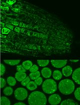
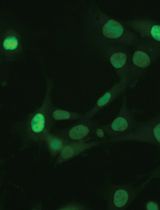
- Assessment of biofilm formation on catheters
- 12-well polystyrene microplate (Fisher Scientific, catalog number: 08-772-50)
- Aluminum foil
- 10% Neutral Buffered Formalin (Fisher Scientific, catalog number: 22-046-361)
- 20% Sodium azide solution (Sigma-Aldrich, catalog number: S-2002)
- Tween-20 (Sigma-Aldrich, catalog number: P1379)
- Methyl α-d-mannopyronoside (Sigma-Aldrich, catalog number: M6882)
- Rabbit anti-Enterococcus antibody (Abcam, catalog number: ab68540) or any other anti-Enterococcus antibody commercially available
- IRDye 680LT goat anti-rabbit (LI-COR Biosciences, catalog number: 926-68021)
- Immunostaining Solutions (see Recipes)
- Blocking Solution
- Wash Solution
- Dilution Buffer
- Primary antibody solution
- Secondary antibody solution
- Assessment of Biofilm on microplate
- Paper towel
- Reagent reservoir (Fisher Scientific, catalog number: 14-387-065)
- Crystal violet (Sigma-Aldrich, catalog number: C0775)
- Acetic acid (Sigma-Aldrich, catalog number: A6283)
- Solutions (see Recipes)
- 0.5% Crystal violet solution
- 33% of Acetic Acid
Equipment
- Forceps
- Heraeus Multifuge X3R Refrigerated Centrifuge (VWR, catalog number: 75004516) or any refrigerated centrifuge with similar features
- Spectra Max ABS Plus Spectrophotometer (Molecular Devices, catalog number: ABS PLUS) or any spectrophotometer with similar features
- Branson UltrasonicsTM BransonicTM CPX-sonicator (waterbath sonicator) (Fisher Scientific, catalog number: 15-337-419) or any waterbath sonicator with similar features
- Odyssey CLx imaging system (LI-COR, catalog number: 9140-01)
- Test tube racks (Fisher Scientific, catalog number: 14-809-62) or any standard test tube racks
- VWR Microbiological Incubator (VWR, catalog number: 51030017) or any standard microbiological incubator
- Biological Safety Cabinet with UV light (Thermo Fisher, catalog number: 1395) or any standard equipment with similar features
- Vortexer (Fisher brand, catalog number: 02-215-418) or any standard vortex
- Autoclave (Getinge, catalog number: 633LS) or any standard autoclave
Software
- CLX image studio (LI-COR, catalog number: 9140-510)
- GraphPad Prism (GraphPad Software LLC)
Procedure
文章信息
版权信息
© 2019 The Authors; exclusive licensee Bio-protocol LLC.
如何引用
Colomer-Winter, C., Lemos, J. A. and Flores-Mireles, A. L. (2019). Biofilm Assays on Fibrinogen-coated Silicone Catheters and 96-well Polystyrene Plates. Bio-protocol 9(6): e3196. DOI: 10.21769/BioProtoc.3196.
分类
微生物学 > 微生物生物膜 > 生物膜培养
生物化学 > 蛋白质 > 免疫检测 > 免疫染色法
分子生物学 > 蛋白质 > 蛋白质-蛋白质相互作用
您对这篇实验方法有问题吗?
在此处发布您的问题,我们将邀请本文作者来回答。同时,我们会将您的问题发布到Bio-protocol Exchange,以便寻求社区成员的帮助。