

High-resolution Immunoelectron Microscopy Techniques for Revealing Distinct Subcellular Type 1 Cannabinoid Receptor Domains in Brain
检测脑中不同亚细胞1型大麻素受体结构域的高分辨率免疫电镜技术

(*contributed equally to this work) 发布: 2019年01月20日第9卷第2期 DOI: 10.21769/BioProtoc.3145 浏览次数: 9750
评审: Edgar Soria-GomezEstibaliz GonzalezAnonymous reviewer(s)
参见作者原研究论文
The authors used this protocol in:

Nov 2017
Advertisement


Abstract
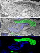
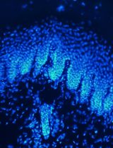
Activation of type 1 cannabinoid (CB1) receptors by endogenous, exogenous (cannabis derivatives) or synthetic cannabinoids (i.e., CP 55.940, Win-2) has a wide variety of behavioral effects due to the presence of CB1 receptors in the brain. In situ hybridization and immunohistochemical techniques have been crucial for defining the CB1 receptor expression and localization at the cellular level. Nevertheless, more advanced methods are needed to reveal the precise topography of CB1 receptors in the brain, especially in unsuspected sites such as other cell types and organelles with low receptor expression (e.g., glutamatergic neurons, astrocytes, mitochondria). High-resolution immunoelectron microscopy provides a more precise detection method for the subcellular localization of CB1 receptors in the brain. Herein, we describe a single pre-embedding immunogold method for electron microscopy based on the use of specific CB1 receptor antibodies and silver-intensified 1.4 nm gold-labeled Fab' fragments, and a combined pre-embedding immunogold and immunoperoxidase method that employs biotinylated secondary antibodies and avidin-biotin-peroxidase complex for the simultaneous localization of CB1 receptors and protein markers of specific brain cells or synapses (e.g., GFAP, GLAST, IBA-1, PSD-95, gephyrin). In addition, a post-embedding immunogold method is also described and compared to the pre-embedding labeling procedure. These methods provide a relatively easy and useful approach for revealing the subcellular localization of low amounts of CB1 receptors in glutamatergic synapses, astrocytes, neuronal and astrocytic mitochondria in the brain.
Keywords: Endocannabinoid system (内源性大麻素系统)Background
The endocannabinoid system (eCBs) is widely distributed in the nervous system, and, through the activation of CB1 receptors, plays an important role in normal brain function (Herkenham et al., 1990; Tsou et al., 1998; Kano et al., 2009; Castillo, 2012; Katona and Freund, 2012; Lutz et al., 2015; Pertwee, 2015; Lu and Mackie, 2016; Busquets-Garcia et al., 2018).
Pre- and post-embedding immunocytochemical techniques for electron microscopy are valuable methods that can provide precise CB1 receptor localization in brain and peripheral tissues. The pre-embedding immunogold labeling method has been successfully employed in our laboratory for the localization of various receptors, ion channels and enzymes in the mammalian brain, and has served to reveal a unique and interesting presence of CB1 receptors in mitochondria (Bénard et al., 2012; Hebert-Chatelain et al., 2014 and 2016; Mendizabal-Zubiaga et al., 2016; Gutiérrez-Rodríguez et al., 2018). The advantages are reflected by its ease of applicability and relatively inexpensive tissue preparation equipment and relatively good ultrastructural preservation. In addition, it offers the ability to perform correlative light microscopy on stained sections in order to reveal general patterns of CB1 receptor labeling prior to examination with the electron microscope. Success of the pre-embedding detection method has been improved by the development of ultra-small gold secondary conjugates (Nanogold®) in combination with gold particle enlarging chemistry, making it possible for probes to detect deeper into tissues, thus reducing the inherent limitation of antibody penetration and molecule detection while enhancing the resolution of receptor localization. In combination with immunoperoxidase and 3,3’-diaminobenzidine (DAB) reaction product immunochemistry, pre-embedding procedures can reveal protein co-localizations with cell specificity and at high resolution.
With post-embedding immunogold labeling methods, the epitopes are theoretically exposed at the section surface, diffusion of immunoreagents is avoided, is quantitative, allows simultaneous labeling by using different gold particle sizes (Hermida et al., 2010, Hunt et al., 2013) and the labeling is reliable on serial ultrathin sections (Hermida et al., 2006). However, the sensitivity is only moderate, not all antibodies work successfully in the resin-embedded tissue conditions required for the technique, and membrane and structural protein visibility is typically compromised since post-fixation with osmium tetroxide is avoided due to its destructive effects on antigens. Last, but not least, the equipment required for post-embedding labeling procedures can be unaffordable.
Materials and Reagents
- Glass vials (Thermo Fisher Scientific, catalog number: C4010-LV1)
- Glass slides (Sigma-Aldrich, catalog number: S8902)
- Aluminum foil (Sigma-Aldrich, catalog number: 326852)
- Syringes (Proquinorte)
- Nickel mesh grids: grids for pre-embedding electron microscopy (Electron Microscopy Sciences, catalog number: G-150 Ni), storage temperature: RT
- Nickel single slot formvar coated grids (aperture grids: grids for post-embedding electron microscopy) (Electron Microscopy Sciences, catalog number: FFGA1000-Ni-50), storage temperature: RT
- Antibodies
Antibody Manufacturer; species; catalog number; RRID Anti-cannabinoid receptor type-1 (CB1) Frontier Institute Co., ltd; goat polyclonal; CB1-Go-Af450; AB_2571592 Anti-cannabinoid receptor type-1 (CB1) Frontier Institute Co., ltd; guinea pig polyclonal; CB1-GP-Af530; AB_2571593 Anti-glial fibrillary acidic protein (GFAP) Sigma-Aldrich; mouse monoclonal; G3893; AB_257130 Anti-gephyrin Synaptic Systems; mouse monoclonal; 147021; AB_2232546 Anti-A522 (EAAT1 [GLAST]) Prof. Niels Christian Danbolt University of Oslo; rabbit polyclonal; Ab#314; AB_2314561 Anti-metabotropic glutamate receptor 2/3 (mGluR2/3) Chemicon (Millipore); rabbit polyclonal; AB1553, AB_11212089 Biotinylated anti-mouse secondary antibody Vector Labs; BA-2000; AB_2313581 Biotinylated anti-rabbit secondary antibody Vector Labs; BA-1000; AB_2313606 1.4 nm gold-conjugated anti-guinea pig IgG (Fab’ fragment) secondary antibody Nanoprobes; goat; #2055 1.4 nm gold-conjugated anti-goat IgG (Fab’ fragment) antibody Nanoprobes; rabbit; #2004 Colloidal gold-18 nm anti-goat IgG antibody Jackson Immunoresearch; donkey; 705-215-147 F(ab’)2 anti-rabbit IgG fragments conjugated to 10 nm colloidal gold particles British Biocell International; goat; GFAR10 - Lowicryl HM20 kit (Sigma-Aldrich, catalog number: 15924), storage temperature: RT
- VECTASTAIN Elite ABC HRP Kit (Peroxidase, Standard) (Vector Laboratories, catalog number: PK-6100), storage temperature:
4 °C - HQ Silver: silver enhancement kit for EM (Nanoprobes, catalog number: 2012), storage temperature: -20 °C
- Bovine serum albumin (BSA) (Sigma-Aldrich, catalog number: A7906), storage temperature: 4 °C
- 3,3′-diaminobenzidine tetrahydrochloride hydrate (DAB): C12H14N4•4HCl•xH2O (Sigma-Aldrich, catalog number: D5637), storage temperature: -20 °C
- Ethanol absolute: CH3CH2OH (PanReac AppliChem, catalog number: A1613), storage temperature: RT
- Glycerol: HOCH2CH(OH)CH2OH (Sigma-Aldrich, catalog number: G9012), storage temperature: RT
- Glycine: NH2CH2COOH (Sigma-Aldrich, catalog number: G8898), storage temperature: RT
- Ketamine hydrochloride/xylazine hydrochloride solution for anesthesia (Sigma-Aldrich, catalog number: K4138), storage temperature: 4 °C
- Methanol: CH3OH (Sigma-Aldrich, catalog number: 322415), storage temperature: RT
- Propane: CH3CH2CH3 (Sigma-Aldrich, catalog number: 536172), storage temperature: RT
- (R)-(+)-propylene oxide: C3H6O (Sigma-Aldrich, catalog number: 540048), storage temperature: RT
- Saponin (Sigma-Aldrich, catalog number: 84510), storage temperature: RT
- Sodium azide (NaN3) (PanReac AppliChem, catalog number: 122712.1609), storage temperature: RT
- Sodium borohydride: NaBH4 (Sigma-Aldrich, catalog number: 71320), storage temperature: RT
- Osmium tetroxide aqueous solution (4%) (Electron Microscopy Sciences, catalog number: 19150), storage temperature: RT
- Di-Sodium hydrogen phosphate (Na2HPO4) (Merck, catalog number: 1.06586.1000), storage temperature: room temperature (RT) (used in Recipes 1-3)
- Epoxy embedding medium, hardener DDSA: C16H26O3 (Sigma-Aldrich, catalog number: 45346), storage temperature: RT (used in Recipe 6)
- Epoxy embedding medium, hardener MNA: C10H10O3 (Sigma-Aldrich, catalog number: 45347), storage temperature: RT (used in Recipe 6)
- Epoxy embedding medium: EponTM 812 substitute (Sigma-Aldrich, catalog number: 45345), storage temperature: RT (used in Recipe 6)
- Glutaraldehyde (25%) in aqueous solution for synthesis: C5H8O2 (Merck, catalog number: 8.20603.1000), storage temperature: 4 °C (used in Recipe 1)
- Hydrochloric acid (HCl) (Sigma-Aldrich, catalog number: H1758), storage temperature: RT (used in Recipe 2)
- Lead(II) nitrate: (PbNO3)2 (PanReac AppliChem, catalog number: 131473), storage temperature: RT (used in Recipe 8)
- N-benzyldimethylamine: C9H13N (Sigma-Aldrich, catalog number: 185582), storage temperature: RT (used in Recipe 6)
- Paraformaldehyde: (CH2O)n (Merck, catalog number: 1.04005.1000), storage temperature: RT (used in Recipe 1)
- Picric acid solution (1.3%) dissolved in H2O (saturated): (O2N)3C6H2OH (Sigma-Aldrich, catalog number: P6744), storage temperature: RT (used in Recipe 1)
- Poly(ethylene glycol) (PEG): C2nH4nH2On+1 (Sigma-Aldrich, catalog number: 202444), storage temperature: RT
- Potassium chloride (KCl) (Sigma-Aldrich, catalog number: P9541), storage temperature: RT (used in Recipe 2)
- Potassium phosphate monobasic (KH2PO4) (Sigma-Aldrich, catalog number: 229806), storage temperature: RT (used in Recipe 2)
- Sodium chloride (NaCl) (PanReac AppliChem, catalog number: 131659.1211), storage temperature: RT (used in Recipes 2-4)
- Sodium hydroxide pellets (PanReac AppliChem, catalog number: 131687.1211), storage temperature: RT (used in Recipe 8)
- Sodium phosphate monobasic monohydrate (NaH2PO4•H2O) (PanReac AppliChem, catalog number: 131965.1211), storage temperature: RT (used in Recipes 1-3)
- Tri-sodium citrate 2-hydrate: Na3C6H5O7•2H2O (Merck, catalog number: 6448), storage temperature: 4 °C (used in Recipe 8)
- Triton X-100 (Sigma-Aldrich, catalog number: X100), storage temperature: RT (used in Recipe 5)
- Trizma® base: NH2C(CH2OH)3 (Sigma-Aldrich, catalog number: T1503), storage temperature: RT (used in Recipes 4 and 5)
- Trizma® hydrochloride: NH2C(CH2OH)3•HCl (Sigma-Aldrich, catalog number: T3253), storage temperature: RT (used in Recipe 4)
- Uranyl acetate: UO2(CH3COO)2 (Electron Microscopy Sciences, catalog number: 22400), storage temperature: RT (used in Recipe 7)
- Fixative solution (see Recipes)
- Phosphate buffered saline (PBS 1x) (see Recipes)
- 0.1 M phosphate buffer (0.1 M PB) (see Recipes)
- Tris-hydrogen chloride buffered saline (TBS 1x) (see Recipes)
- Tris-buffered saline with Triton X-100 (TBST) (see Recipes)
- Epon resin (see Recipes)
- Uranyl acetate (2%, aqueous) staining solution (see Recipes)
- Reynold’s lead citrate staining solution (see Recipes)
Equipment
- Erlenmeyer flasks (Sigma-Aldrich, catalog number: Z567868)
- Shaker (Heidolph, Duomax 1030)
- Oven (Grupo Selecta, Dryterm)
- Vibratome (Leica Biosystems, model: Leica VT1000 S)
- Super platinum knife (Gillette)
- Ultramicrotome (RMC Products, model: PowerTome XL)
- Histo 6 mm diamond knife 45° (Diatome)
- Ultra 2.4 mm diamond knife 45° (Diatome)
- Transmission electron microscope (Philips, model: EM208S)
- Digital Morada camera (Olympus SIS Morada camera)
- Cryofixation unit (Reichert-Jung, model: KF 80)
- Freeze substitution system (Reichert AFS)
Software
- Adobe Photoshop (CS3, Adobe Systems, San Jose, CA, USA)
- ImageJ (NIH, USA; RRID:SCR_003070)
- GraphPad Prism 5 (GraphPad Software Inc., San Diego, USA; RRID:SCR_002798)
Procedure
文章信息
版权信息
© 2019 The Authors; exclusive licensee Bio-protocol LLC.
如何引用
Puente, N., Bonilla-Del Río, I., Achicallende, S., Nahirney, P. C. and Grandes, P. (2019). High-resolution Immunoelectron Microscopy Techniques for Revealing Distinct Subcellular Type 1 Cannabinoid Receptor Domains in Brain. Bio-protocol 9(2): e3145. DOI: 10.21769/BioProtoc.3145.
分类
神经科学 > 神经解剖学和神经环路 > 脑神经
发育生物学 > 细胞信号传导 > G蛋白偶联受体
细胞生物学 > 细胞染色 > 蛋白质
您对这篇实验方法有问题吗?
在此处发布您的问题,我们将邀请本文作者来回答。同时,我们会将您的问题发布到Bio-protocol Exchange,以便寻求社区成员的帮助。