

Microirradiation for Precise, Double-strand Break Induction in vivo in Caenorhabditis elegans
显微照射用于秀丽隐杆线虫中双链断裂的精确体外诱导
发布: 2018年12月20日第8卷第24期 DOI: 10.21769/BioProtoc.3130 浏览次数: 5288
评审: Neelanjan BoseLivia UlicnaJuan Facundo Rodriguez Ayala
参见作者原研究论文
The authors used this protocol in:

Jan 2018
Advertisement


Abstract
DNA double-strand breaks (DSBs) are toxic lesions that every cell must accurately repair in order to survive. The repair of DSBs is an integral part of a cell life cycle and can lead to lethality if repaired incorrectly. Laser microirradiation is an established technique which has been used in yeast, mammalian cell culture, and Drosophila cell culture to study the regulation of DSB repair. Up to our studies, this method has not been adapted for use in a whole, live, multicellular organism to study this repair in vivo. We have recently shown that this system can be used for study of the recruitment of vital repair proteins to microirradiation-induced breaks in the transparent nematode Caenorhabditis elegans. With the integration of microirradiation and imaging technology, we can precisely induce DSBs in target nuclei and study the recruitment of fluorescently tagged repair proteins from the time of damage induction. Whole, live worms are plated and immobilized for targeting of nuclei, and immediately following induction the targeted region can be imaged for up to an hour and a half post-microirradiation. This method is the first that allows for study of DNA repair protein kinetics in vivo in an intact organism, which can be adapted in numerous ways to allow for study of repair kinetics in various aspects of the repair process.
Keywords: Microirradiation (显微照射)Background
DNA double-strand breaks (DSBs) are one of the most toxic forms of DNA damage and can be induced exogenously (e.g., UV damage) or endogenously (e.g., SPO-11-induced meiotic DSBs). Studying the recruitment of proteins to the sites of DSBs provides valuable information regarding how the process, and specific proteins involved, are regulated. Microirradiation has been extensively used in cell culture as a means of studying DNA repair (Aten et al., 2004; Kong et al., 2009). This method, coupled with fluorescently-labeled proteins and time-lapse imaging, has provided critical information regarding the regulation of proteins in specific cellular contexts. However, this incredibly tractable method had not been adapted for use in a whole, live, multicellular organism.
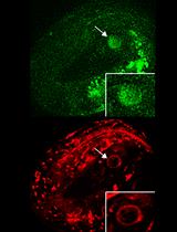
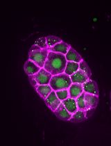
In our paper, we describe a laser microirradiation method which applies this technology to intact, live worms (Koury et al., 2018). With the use of a UVA 365 nm pulsed laser, and fluorescently tagged repair proteins RPA-1 and RAD-51, we were able to precisely induce DSBs in specific regions of the C. elegans germline to study DSB repair kinetics in meiotic tissue in vivo. RPA-1 and RAD-51 are ssDNA binding proteins that are essential for DSB repair using homologous recombination. The transparent nature of worms allows for live imaging of fluorescent proteins without any form of dissection or gonad extrusion, and no pre-sensitization of the worms is required for efficient damage induction. Not only is this method straight-forward, but it can be adapted for, and applied to, numerous questions in the field of DSB repair and regulation in both meiotic and somatic tissues.
Materials and Reagents
Note: We indicated our vendors and cat numbers for lab reagents such as tips, tubes, and chemicals, however, any vendor will likely be suitable and sufficient.
- 0.2-20 μl Pipette tips (VWR, catalog number: 89079-438)
- Microscope slides, 1” x 3” x 0.4” (Surgipath-Leica, catalog number: 3800240)
- Petri dishes, any 60 x 15 mm plate with ventilation ribs (Kord-Valmark, catalog number: 2901)
- Glass bottom dishes: 35 mm dish with 14 mm glass microwell (No. 1.5 coverglass, MatTek, catalog number: P35G-0.170-14-C)
- Parafilm, 4” x 125’ (Parafilm M, catalog number: PM996)
- Pasteur pipet, 5 ¾” Flint Glass (Fisher Scientific, catalog number: 50-930-565)
- General-Purpose Laboratory Labeling Tape, ¾” (VWR, catalog number: 89098-004)
- 12 mm tube (any type of tube with a 12 mm diameter would work; VWR, catalog number: 20170-579)
- Worm pick, hand-made by attaching platinum wire to a Pasteur pipette using fire
- 0.1-μm polystyrene beads (Polybead, Polysciences, catalog number: 00876, store at 4 °C)
- Dissection blade (Stainless Steel Surgical Blades, 4-311)
- Escherichia coli OP50 (from the Caenorhabditis Genetics Center)
- Agarose (any brand of Agarose that is typically used for gel electrophoresis, e.g., VWR, catalog number: 9012-36-6)
- Immersion oil, 100x/1.4 NA (Leica Microsystems, catalog number: 11513859)
- Sodium chloride (NaCl) (RPI, catalog number: 7647-14-5)
- Peptone (RPI, catalog number: 73049-73-7)
- Cholesterol (Sigma-Aldrich, catalog number: 57-88-5)
- Calcium chloride (CaCl2) (Sigma-Aldrich, catalog number: 10035-04-8)
- Magnesium sulfate (MgSO4) (Sigma-Aldrich, catalog number: 7487-88-9)
- Agar (RPI, catalog number: 9002-18-0)
- Potassium dihydrogen phosphate (KH2PO4) (RPI, catalog number: 7778-77-0)
- Sodium phosphate dibasic (Na2HPO4) (Sigma-Aldrich, catalog number: 7558-80-7)
- Ethanol
- Nematode growth medium (NGM) agar plate (see Recipes)
- M9 buffer (see Recipes)
- 10% agarose pads (see Recipes)
Equipment
- Microscope suitable for live imaging (such as Leica DMi8) with Andor MicroPoint 365 nm pulsed laser (Leica, model: Leica DMi8)
- Humidity chamber (box with wet paper towels and elevated surface)
- Microwave (any brand should work, we use Panasonic Inverter, model: NN-S543BFR)
- Stereo microscope (Leica Microsystems, model: KL200 LED)
- 20 °C incubator (Nor-Lake Scientific Refrigerated Incubator, LRI201WWW/0)
- 37 °C incubator (Precision Scientific Thelco Incubator, model: 31483)
- Autoclave
- 4 °C refrigerator
Software
- MetaMorph Software (Molecular Devices, LLC, version 7.8.12.0)
- Fiji (Fiji is just ImageJ) (Free to download at imagej.net/Fiji) (Schindelin et al., 2012)
- GraphPad Prism v6 (GraphPad Software, La Jolla California USA, www.graphpad.com/)
Procedure
文章信息
版权信息
© 2018 The Authors; exclusive licensee Bio-protocol LLC.
如何引用
Harrell, K. E., Koury, E. and Smolikove, S. (2018). Microirradiation for Precise, Double-strand Break Induction in vivo in Caenorhabditis elegans. Bio-protocol 8(24): e3130. DOI: 10.21769/BioProtoc.3130.
分类
细胞生物学 > 细胞成像 > 活细胞成像
您对这篇实验方法有问题吗?
在此处发布您的问题,我们将邀请本文作者来回答。同时,我们会将您的问题发布到Bio-protocol Exchange,以便寻求社区成员的帮助。