

Partial Transection of Adult Rat Optic Nerve as a Model of Secondary Degeneration in the Central Nervous System
成年大鼠视神经不完全横切作为中枢神经系统继发性损害模型


发布: 2018年12月20日第8卷第24期 DOI: 10.21769/BioProtoc.3118 浏览次数: 5625
评审: Oneil G. BhalalaHong LianKarthik Krishnamurthy
参见作者原研究论文
The authors used this protocol in:

Jul 2018
Advertisement


Abstract
Injury to the central nervous system is characterized by damage that spreads from the initial point of impact into the surrounding adjacent tissue, in a phenomenon referred to as secondary degeneration. The optic nerve can be used to effectively model injury and secondary degeneration to white matter tracts. Partial transection of the dorsal aspect of the nerve leaves the ventral aspect initially undamaged but vulnerable to secondary degeneration, allowing study of tissue exclusively vulnerable to secondary degeneration. Thus the partial optic nerve transection model of secondary degeneration is a valuable tool to study the pathology of spreading damage following neurotrauma and can be used to assess potential efficacy of therapeutic strategies.
Keywords: Neurotrauma (神经损伤)Background
Traumatic injury to the central nervous system (CNS) is the direct damage to the brain or spinal cord resulting from a physical insult. Traumatic brain injury and spinal cord injury are major public health issues and have significant associated costs (Access Economics, 2009). The optic nerve has long been used as a model for CNS injury, particularly to white matter tracts, and the response to injury has been well characterized in a range of species, from fish where there is complete regeneration of severed retinal ganglion cell axons, to mammals where regeneration is abortive, and damage is compounded by the process of secondary degeneration (Harvey et al., 2006). As a model, the optic nerve has many attractive features: it is a white matter tract that is accessible, the level of damage can be easily controlled, the associated neuronal cell bodies are in the eye and can easily be studied, downstream anatomical pathways are well described and the impact of damage or potential therapeutic intervention can be studied at the molecular, biochemical, histological and behavioral levels. As such, damage to the optic nerve can be used to study neurotrauma, secondary degeneration, regeneration, neuronal plasticity, and remote responses.
Commonly injury to the CNS is partial, with a focal insult and adjacent initially spared tissue. The trauma triggers a cascade of secondary events, creating a toxic environment in the adjacent tissue and leading to progressive secondary degeneration with increasingly widespread pathology, as well as tissue and functional loss over time. Therapeutic targeting of secondary degeneration is possibly the most promising avenue for treatment of CNS trauma, as it involves protection of initially undamaged tissue rather than repair of transected neurons (Fitzgerald, 2014). However, the mechanisms by which these secondary events occur are complex and need to be elucidated in order to develop treatment strategies. Resultant therapeutics may be relevant to other conditions such as stroke and glaucoma, where similar mechanisms may apply (Tezel, 2006). Partial transection of the optic nerve in rat provides an excellent model to study secondary degeneration (Levkovitch-Verbin et al., 2003; Fitzgerald et al., 2009). This is because, in contrast to optic nerve crush injuries, only retinal ganglion cell (RGC) axons in the dorsal aspect of the nerve are transected, allowing spatial segregation of a primary injury from adjacent tissue in ventral nerve that is exclusively subject to secondary degeneration (Payne et al., 2012). Additionally, RGCs in ventral retina are subject only to secondary degenerative events whilst those in dorsal retina are subject to both primary and secondary insults (Payne et al., 2012). Thus, the partial transection model is particularly useful for examining events associated with secondary degeneration, since there is a clear delineation between axons in dorsal nerve that are transected by the primary injury and axons lying ventrally that are initially spared but are affected by secondary degenerative events. This protocol describes in detail the surgical and associated procedures used to perform a partial transection of the optic nerve in adult rats. The procedure is best performed with two people, one to do the pre- and post-operative care and one to do the surgery.
Materials and Reagents
Note: Unless otherwise stated, items are supplied by general laboratory suppliers and veterinary or medical suppliers.
- Sterilized items (autoclaved or purchased as sterile)
- 1 ml syringes (Becton Dickenson, catalog number: 309659)
- Gauge 27 needles, 3 per animal (Becton Dickenson, catalog number: 305109)
- Cotton wool balls (Supermarket home brand)
- Surgical gauze swab, cut into 1cm squares (ProVet, GAUZ HS N1)
- Small animal surgical drapes with window of approximately 2 cm x 4 cm (Theatrewrap, SA)
- Silicon instrument mat (e.g., Medline DYNJHOLD1)
- Square restaurant grade linen napkin(s) (e.g., Lunaweddingandeventsupplies.com.au, black linen napkin 50 cm x 50 cm)
- Stainless steel instrument dish (e.g., Instrumentation concept, KDY8)
- Scalpel blade #11 (ProVet, SCAL HS11) (or to your preference)
- Suture thread [Silkam #4 (AMA, BRAC0762130) or #6 (AMA,BRAC0762113) or to your preference]
- Surgical gloves (ProVet, GLOV CLF7)
- Distilled water (in house, autoclaved in Schott bottles)
- Phosphate buffered saline pH 7.2 (PBS) (in house, autoclaved in Schott bottles)
- Non-sterile items
- Permanent marker (Officeworks)
- Spray bottles containing 70% ethanol (Spray bottle from hardware store, e.g., Bunnings)
- Clean gown (ProVet, GOWN S KS)
- Hair cover (e.g., Medline NONBOUF21)
- Surgical mask (ProVet, SURG MHS03)
- Autoclave bags (Provet, Henry Schen, various sizes, AUTO P HM2; AUTO P HM4)
- Bubble wrap cut to size to place around the rat (Officeworks)
- Tape (masking tape, Officeworks)
- Paper towel (Tork)
- Tube rack (Thermo Fisher, 21-200-285)
- 50 ml Falcon tube (Thermo Fisher, NUN339652) containing 70% ethanol
- Small Petri dishes (Thermo Fisher, Sterilin 121V)
- 70% ethanol
- Vaseline
- Biological material
Rats [Female Piebald Viral Glaxo (PVG) rats 160 g+ obtained from Animal Resource Centre (ARC), Canning Vale, WA 6970] - Drugs
Note: All drugs purchased through ProVet (provet.com.au).- Ketamine [Ketamil, Troy Laboratories, 50 mg/kg [50 mg/ml] diluted in phosphate buffered saline (PBS)]
Note: Ketamil is a scheduled poison and should be stored with appropriate levels of security. It should be protected from light and stored at room temperature, observing expiry date on the product label. - Xylazil [Xylazil, Troy Laboratories, 10 mg/kg (10 mg/ml) diluted in PBS (Store at 20-25 °C and observe product expiry date on label.)]
- Carprieve (Norbrook Australia, Pty. Ltd.; Victoria, Australia, 5 mg/kg (5 mg/ml in sterile water) (Store protected from light at 2 °C to 8 °C. Once open store refrigerated for up to 28 days.)
- Luxyl protective eye drops (Tubilux Pharma SpA Via Costarica 20/22 Pomezia Italy) (Store below 25 °C. Once open store refrigerated for a maximum of 60 days and dispose of immediately should sterility be compromised.)
- Betadine (500 ml ProVet, BETA AS) (Store below 25 °C protected from light, observing any expiry information on the label.)
- Ketamine [Ketamil, Troy Laboratories, 50 mg/kg [50 mg/ml] diluted in phosphate buffered saline (PBS)]
Equipment
- Schott bottles 50-250 ml
- Surgical instruments (Kaisers surgical instruments Pty Ltd)
- Watchmakers forceps (one or two coarse, one or two medium and two fine points)
- A medium pair with tips bent inwards towards each other to form a ‘hook’ is also useful
- Fine dissecting spring-form scissors (in different sizes)
- Needle holder or small Spencer Wells forceps for suturing
- Small scissors for cutting suture thread
- Bench-top autoclave (Siltex, Practica 20L lab steam sterilizer)
- Weigh balance suitable for weighing animals (e.g., Mettler Toledo, MW 1002T/00)
- Small animal clippers (e.g., Wahl KM2)
- Binocular surgical/dissecting microscope with sufficient working distance (e.g., Nikon, model: SMZ460)
- Light source to illuminate surgical site (e.g., Schott, catalog number: KL 1500 LED)
- Clear Perspex animal supports approx. 25 x 10 cm (made in house; also available from Plastic Warehouse)
- Mouth bar (Made in house through engineering workshop, Figure 1) (An alternative would be to adapt the mouth bar from a stereotaxic frame such as the WPI 502612)
- Small retort stand and boss head clamp (Labdirect, Kartell retort stand; Technos boss head clamp)
- Diamond keratotomy knife (Geuder, catalog number: G-19345)
- Small animal heat pad (My pet warehouse, Ref 234016)
- Spare animal cage(s) for use during induction of anesthesia and/or after recovery
- Anal thermometer
Note: Also required is a suitable surgical table or bench space.
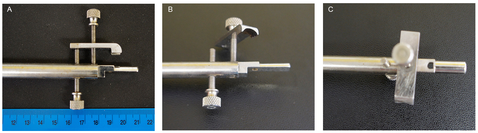
Figure 1. Stainless steel mouth bar. A. The mouth bar was machined from a stainless steel rod. (B) Shaped to slip into the mouth and (C) with a hole for the top incisors to go through. B and C. The nose clamp has a curved end to accommodate the nose and is tightened with the use of two screws. One presses the clamp onto the nose and the second applies slight upwards pressure from beneath the nose clamp.
Procedure
文章信息
版权信息
© 2018 The Authors; exclusive licensee Bio-protocol LLC.
如何引用
Readers should cite both the Bio-protocol article and the original research article where this protocol was used:
- Bartlett, C. A. and Fitzgerald, M. (2018). Partial Transection of Adult Rat Optic Nerve as a Model of Secondary Degeneration in the Central Nervous System. Bio-protocol 8(24): e3118. DOI: 10.21769/BioProtoc.3118.
- Giacci, M. K., Bartlett, C. A., Smith, N. M., Iyer, K. S., Toomey, L. M., Jiang, H., Guagliardo, P., Kilburn, M. R. and Fitzgerald, M. (2018). Oligodendroglia are particularly vulnerable to oxidative damage after neurotrauma in vivo. J Neurosci 38(29): 6491-6504.
分类
神经科学 > 神经系统疾病 > 动物模型
细胞生物学 > 组织分析 > 损伤模型
您对这篇实验方法有问题吗?
在此处发布您的问题,我们将邀请本文作者来回答。同时,我们会将您的问题发布到Bio-protocol Exchange,以便寻求社区成员的帮助。