

Systematic Quantification of GFP-tagged Protein Foci in Schizosaccharomyces pombe Nuclei
粟酒裂殖酵母细胞核中GFP标记蛋白聚集点的系统量化

发布: 2018年12月20日第8卷第24期 DOI: 10.21769/BioProtoc.3117 浏览次数: 6127
评审: Dennis NürnbergGunjan MehtaSamantha E. R. Dundon
参见作者原研究论文
The authors used this protocol in:
Jun 2018
Advertisement


Abstract
DNA damage repair proteins form foci in response to DNA damaging agents. The efficiency and integrity of the DNA repair pathway of a particular eukaryotic (mutant) strain is usually determined by the number of foci formed compared with their wild-type counterpart. Conventionally, focus number is determined visually, and this low accuracy may obscure the identification of a weaker phenotype, particularly when the output is low. Here, using the homologous recombination protein Rhp54 as an example, we present a protocol that can increase the consistency of foci identification among samples and can significantly improve the efficiency of foci quantification for large sample sizes. A similar method can be applied to other foci-forming proteins.
Keywords: Fission yeast (裂殖酵母)Background
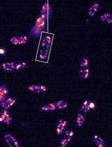
Eukaryotic cells are equipped with a set of DNA damage response mechanisms to defend themselves against internal and external stresses. External stresses, such as chemical agents cause damage or mutation to the DNA and, if left unrepaired, will propagate to the offspring and lead to genomic instability. Methyl methanesulfonate (MMS) is one example of a chemical (alkylating) agent that methylates DNA bases and stalls the replication fork, thereby generating double-strand breaks (DSBs) when the replication fork collapses (Arnaudeau et al., 2001). In Schizosaccharomyces pombe, DSBs are often repaired by the error-free homologous recombination (HR) pathway (Ferreira and Cooper, 2004). HR proteins, such as Rhp54, lead to foci formation in response to MMS treatment, with the number of foci formed increasing proportionately to the level of DNA damage (Maki et al., 2011). Recent work has shown that a mutation in the histone H3 methyltransferase Set2 can affect Rhp54 foci formation during MMS treatment, thereby suggesting a role for Set2 in MMS resistance (Lim et al., 2018).
Quantification of foci number can reflect the robustness of DNA repair mechanisms in cells. However, measuring foci formation efficiency using fluorescently tagged DNA repair proteins often involves manual counting, which is susceptible to human error and is time-consuming (Rübe et al., 2008; Lorenz et al., 2009). There are several free or commercially available software on the market that afford better accuracy in foci determination and increase the throughput; however, there are several problems with such software (Herbert et al., 2014; Ledesma-Fernández and Thorpe, 2015; Oeck et al., 2015; Wiesmann et al., 2015). First, there is lack of a fully systematic and user-friendly way to move from image acquisition to image processing to foci analysis (counting); indeed, a user-friendly program must allow the user to have more control during each step of the process without requiring a background in computer programming (scripts and macros). Second, most of the available software or plugins were developed based on foci formation in mammalian cells, particularly targeting γH2AX foci, and thus might not be suitable for determining foci formation in yeast cells, which are much smaller in size (Böcker and Iliakis, 2006; Jucha et al., 2010; Anderson et al., 2013). The FociQuant software has been used for foci quantification in yeast with few complications; this is because the protocol focuses on the kinetochore protein, which forms a single distinct focus due to the clustering of centromeres (Ledesma-Fernández and Thorpe, 2015). However, difficulties arise in the quantification of foci for HR proteins; because DSBs can occur anywhere in the genome, multiple foci can be scattered in the nucleus and in different focal planes. Therefore, we are unable to use pre-existing methods of foci quantification to determine the foci of HR proteins accurately.
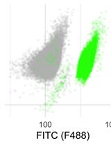
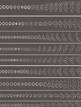
To overcome these problems, we employed the Nikon microscope system for image acquisition and NIS-Elements Advance Research software (v5.02) with the Extended Depth of Focus (EDF) plugin and “General Analysis” for image processing and foci analysis, respectively. Similar to other advanced microscope systems, DAPI and GFP Z-stack images are captured in a single file with one mouse-click. Because the cells exist in a 3D conformation, the EDF function enables the focused image of each plane captured by the Z-stack to be combined into a single, focused image. In this way, foci at different focal planes are included, thereby increasing the accuracy of the quantification. Furthermore, the software enables automated foci quantification by first determining the nucleus via DAPI staining, followed by counting the number of GFP foci in it; this prevents the inclusion of other non-nucleus, GFP noise. Although the analysis process is automated, the user can manually deselect regions or noise that are incorrectly determined by the software. The control and commands on the software are user-friendly, and the user does not need to have programming knowledge to modify certain quantification steps. In addition, the software is not only suitable for the analysis of images captured by a Nikon microscope but can be used to analyze images taken from other sources through additional steps.
Here, using MMS-dependent activation and foci formation of GFP-tagged Rhp54 as an example, we present an unbiased and precise method for the quantification of foci number formed by Rhp54-GFP in yeast. We have applied this method in recent work (Lim et al., 2018) to count foci number in wild-type and set2-mutant cells following MMS treatment.
Materials and Reagents
- Pipette tips
- Microcentrifuge tubes (1.5 ml) (Eppendorf, catalog number: 0030120086)
- Centrifuge tubes (Sigma-Aldrich, model: Greiner 15 ml, catalog number: T1818)
- Microscope slides (Continental Lab Products, catalog number: 7105)
- Microscope cover glasses 22 x 22 mm (Paul Marienfeld GmbH & Co, Marienfeld SUPERIOR, catalog number: 0101050)
- Grade GF/C Glass microfiber filters, 25 mm circle, 1.2 μm pore size (Whatman, catalog number: 1822-025)
- C-terminal GFP-tagged Rhp54 S. pombe strain (Tang et al., 2015; Lim et al., 2018)
- 972, S. pombe wild-type (WT) strain (without any GFP tagging) (Lim et al., 2018)
- Methyl methanesulfonate (MMS) in 99% stock (Sigma-Aldrich, catalog number: 129925)
CAUTION: MMS is a toxic substance and a potential carcinogen. Users should wear all protective wears while working with it. MMS-containing waste media should be placed in a container for cytotoxic waste before disposal by a licensed chemical waste disposal company. - Methanol (AR-grade) in 100% (Fisher Scientific, catalog number: 10284580)
- 4',6-diamidino-2-phenylindole (DAPI) for nucleus staining (Sigma-Aldrich, catalog number: D8417)
- NaCl (Sigma-Aldrich, catalog number: S9888)
- KCl (Sigma-Aldrich, catalog number: P5405)
- Na2HPO4 (Sigma-Aldrich, catalog number: S5136)
- KH2PO4 (Sigma-Aldrich, catalog number: P9791)
- Hydrochloric acid 37% (Sigma-Aldrich, catalog number: 320331)
- Yeast extract (BD Biosciences, BactoTM, catalog number: 212750)
- Adenine (Sigma-Aldrich, catalog number: A8626)
- Glucose (Sigma-Aldrich, catalog number: G8270)
- Immersion Oil for microscopy (Nikon, catalog number: MXA20235)
- 1x phosphate-buffered saline (PBS) (see Recipes)
- 2 N Hydrochloric acid (HCl) (see Recipes)
- Yeast extract adenine (YEA) media for cell growth (see Recipes)
Equipment
- P20 Pipetman (Gilson, catalog number: F123600)
- P200 Pipetman (Gilson, catalog number: F123601)
- P1000 Pipetman (Gilson, catalog number: F123602)
- Conical flask, 250 ml (Kartell, catalog number: 1462)
- Shaker incubator (Eppendorf, model: Innova® 44, catalog number: M1282-0002)
- Inverted fluorescence microscope (e.g., Nikon; model: Eclipse Ti-E) with:
- Oil immersion objective lens (Nikon; model: CFI PLAN APO VC 100XH 1.4 NA)
- DAPI filter (excitation λ: 350 nm; emission λ: 460 nm) (Chroma; model: ET-DAPI; catalog number: 49000)
- FITC filter (excitation λ: 480 nm; emission λ: 535 nm) (Chroma; model: FITC /EGFP /Bodipy FI /Fluo3 /DiO; catalog number: 41001)
- Digital camera (Hamamatsu; model: ORCA-Flash2.8; catalog number: C11440-10C)
- Low-temperature freezer (-80 °C) (Thermo Fisher Scientific, model: FormaTM 700 Series)
- Refrigerated centrifuge (4 °C) (TOMY DIGITAL BIOLOGY, model: MX-305)
- Forceps (Sigma-Aldrich, catalog number: F4267)
- Spectrophotometer (Biochrom, ULTRASPEC, catalog number: 80-2116-30)
- Vacuum filtration holder set, consisting of a conical collection flask and a filter holder (Fisherbrand, catalog number: 09-753G)
- Vacuum pump (Rocker Scientific, model: Rocker 400, catalog number: 167400-11(22))
- Autoclave (ALP, model: CL-40M)
Software
- Nikon NIS Elements AR (v5.02) with Extended Depth of Focus (EDF) and General Analysis plugin (purchased from Nikon Co.)
- Microsoft Office 365–Excel
Procedure
文章信息
版权信息
© 2018 The Authors; exclusive licensee Bio-protocol LLC.
如何引用
Lim, K. K. and Chen, E. S. (2018). Systematic Quantification of GFP-tagged Protein Foci in Schizosaccharomyces pombe Nuclei. Bio-protocol 8(24): e3117. DOI: 10.21769/BioProtoc.3117.
分类
微生物学 > 微生物细胞生物学 > 细胞成像
细胞生物学 > 细胞成像 > 荧光
您对这篇实验方法有问题吗?
在此处发布您的问题,我们将邀请本文作者来回答。同时,我们会将您的问题发布到Bio-protocol Exchange,以便寻求社区成员的帮助。