

Blinded Visual Scoring of Images Using the Freely-available Software Blinder
使用免费软件Blinder对图像进行盲视觉评分
发布: 2018年12月05日第8卷第23期 DOI: 10.21769/BioProtoc.3103 浏览次数: 6435
评审: Pengpeng LiAnonymous reviewer(s)
参见作者原研究论文
The authors used this protocol in:

May 2018
Advertisement


Abstract
In nearly all subfields of biomedical sciences, there are phenotypes that are currently classified by expert visual scoring. In research applications, these classifications require the experimenter to be blinded to the treatment group in order to avoid unintentional bias in scoring. Currently, many labs either use laborious and tedious methods to manually blind the images, require multiple experimenters to gather and score the data blindly or fail to properly blind the data altogether. In this protocol, we present a simple, freely available software that we created that allows the experimenter to blindly score images. In our protocol, the user loads unblinded images and defines a scoring system. The software then shows the user the images in a random order, allowing the user to select a score from their defined scoring system for each image. Furthermore, the software has an optional “quality control” mechanism where the user will be shown some images multiple times to test the robustness of the visual scoring. Finally, the software summarizes the results in an exportable file that includes unblinded summary data for each group and a full list of images with their scores. In this protocol, we briefly present directions for using the software, potential applications, and caveats/limitations to this approach.
Keywords: Blinder (Blinder)Background
Although considerable efforts are being made to automate image processing and analysis through machine learning and other computational approaches (Gulshan et al., 2016; Janowczyk and Madabhushi, 2016; Esteva et al., 2017; Bychkov et al., 2018), many biomedical subfields currently require expert visual image classification due to the complexity of the phenotypes being studied. Furthermore, machine learning approaches require many examples to train, so developing automated approaches to score newly discovered or rare phenotypes may take time. Examples of expert image scoring include but are not limited to: medical diagnostic scores for tumors and other pathologies (Dhyani et al., 2015; Fuchs et al., 2018), veterinary applications (Barton et al., 2018), and research that requires quantification of physiological and cellular morphologies (Passeri et al., 2009; Bretman et al., 2010; Green et al., 2011; Gonzalez-Hunt et al., 2014; Riley et al., 2016).
Expert image scoring requires that the experimenter be blinded to the treatment group in order to avoid unintentional bias in the qualitative visual scoring. Researchers commonly achieve this in one of three ways. First, some labs choose to have multiple experts involved in the preparation of slides/specimens, image acquisition, and image processing/scoring so that treatment groups are not identifiable between tasks (Bretman et al., 2010; Green et al., 2011). A second approach is to have a single experimenter prepare specimens, acquire images, and score images, but have a second experimenter manually rename images to blind the scoring to treatment group. A third approach is to create an in-house command line script to blind the image names to avoid the manual manipulation of file names (Riley et al., 2016). All three of these approaches ultimately can lead to a robust and unbiased classification of images, but are labor-intensive, time-consuming, and can lead to errors in assignment of scores.
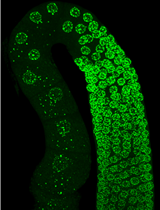
In this protocol, we introduce a simple, freely available software application we developed to allow the user to load unblinded images, be shown those images in a randomized order, easily assign scores, and receive summarized results in a readily exportable file format. All of these steps are carried out within the software using a simple graphical user interface. The software also includes an optional Quality Control mechanism to help identify scoring inconsistencies. We have recently reported exercise effects on mitochondrial morphology in body wall muscle of the nematode Caenorhabditis elegans that were classified by blind visual categorical scoring using this software (Hartman et al., 2018). Herein, we describe the basic methods for using the software as well as caveats and considerations that should be taken into account when using blind visual scoring of images.
Materials and Reagents
- Glass slide
- Coverslip
- Specimen of choice: Caenorhabditis elegans expressing GFP in the mitochondria of body wall muscle (strain SJ4103)
- 10 mM sodium azide (Sigma-Aldrich, St. Louis, MO)
- Agarose
Note: In our example, we used adult Caenorhabditis elegans expressing GFP in the mitochondria of body wall muscle (strain SJ4103); this strain was obtained from the Caenorhabditis Genetics Center (CGC). We immobilized the animals using 10 mM sodium azide (Sigma Aldrich, St. Louis, MO) and mounted them on a 2% agarose pad on a glass slide covered with a coverslip (Shaham, 2006).
Equipment
- LSM 510 confocal microscope (ZEISS, model: LSM 510) or camera to obtain images
Note: In the presented example, we imaged body wall muscle of nematodes using confocal imaging on a Zeiss LSM 510 confocal microscope with a 40x objective.
Software
- Blinder (Solibyte Solutions, Durham, NC; http://blinder.solibytesolutions.com/)–available for Windows operating system only
Note: The software is also archived on Zenodo (DOI: 10.5281/zenodo.1464815). - Image-processing software to prepare images for scoring
FIJI (a distribution of ImageJ https://fiji.sc/) - Statistical software to analyze data
GraphPad Prism 7.04 (https://www.graphpad.com/scientific-software/prism/)
Note: In this protocol, we used FIJI (a distribution of ImageJ) to generate Z-projections of confocal stacks and crop the image to include only body wall muscle from a single animal before loading into the scoring software. We used GraphPad Prism 7.04 to statistically analyze the data from the visual scoring.
Procedure
文章信息
版权信息
© 2018 The Authors; exclusive licensee Bio-protocol LLC.
如何引用
Cothren, S. D., Meyer, J. N. and Hartman, J. H. (2018). Blinded Visual Scoring of Images Using the Freely-available Software Blinder. Bio-protocol 8(23): e3103. DOI: 10.21769/BioProtoc.3103.
分类
细胞生物学 > 细胞成像 > 共聚焦显微镜
您对这篇实验方法有问题吗?
在此处发布您的问题,我们将邀请本文作者来回答。同时,我们会将您的问题发布到Bio-protocol Exchange,以便寻求社区成员的帮助。