

Staining the Germline in Live Caenorhabditis elegans: Overcoming Challenges by Applying a Fluorescent-dye Feeding Strategy
利用荧光染料喂食的方法活体染色标记秀丽隐杆线虫生殖腺

发布: 2018年11月05日第8卷第21期 DOI: 10.21769/BioProtoc.3077 浏览次数: 7293
评审: Neelanjan BoseJuan Facundo Rodriguez AyalaSwati Jalgaonkar
参见作者原研究论文
The authors used this protocol in:
Nov 2017
Advertisement


Abstract
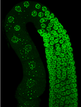
C. elegans provides a tractable model organism for studying germline cell biology. Microscopy experiments are relatively facile, as this worm is transparent and germline development can be observed in real-time using DIC microscopy and/or fluorescent transgenes. Despite these many tools, robust staining techniques for imaging germ cells in live worms have been more elusive, due to the tough outer cuticle of the worm, which impairs staining efficiency. This limitation has restricted the spectrum of probes that can be used to investigate reproductive cell biology in C. elegans. Building on previous approaches, I recently applied a fluorescent-dye feeding strategy to reproducibly label organelles and monitor physiological changes in germlines of living C. elegans. In this approach, fluorescent dyes are initially introduced into the agar plates and bacterial lawns on which worms are subsequently cultured. After worms are grown on the dyed plates, oocytes show staining patterns consistent with verified transgenic markers. Thus, this approach offers an effective solution for labeling difficult-to-stain tissues in live worms, and establishes an entry point for incorporating new probes and sensors into analyses of C. elegans germline biology.
Keywords: C. elegans (秀丽隐杆线虫)Background
Animal reproduction is one of the most fascinating topics in biology. Understanding how germ (reproductive) cells are able to give rise to offspring with each new generation has intrigued scientists, as well as the general public, for centuries. With recent advances in imaging techniques and tools, researchers have been able to observe oocytes (female germ cells) and sperm (male germ cells) in extraordinary detail, revealing fundamental mechanisms that allow germ cells to execute their specialized functions. Model organism research, in particular, has afforded the opportunity to combine live-animal imaging with genetic analyses to tease apart relevant biological pathways important for germ cell regulation. The nematode C. elegans, which exists as a hermaphrodite that continually self-fertilizes throughout young-adulthood (Klass, 1977), presents a powerful experimental system for studying important questions in reproductive biology. Recently, I utilized the C. elegans germline to explore how signs of aging are reversed from one generation to the next (Bohnert and Kenyon, 2017). These studies required the use of dyes to track various aspects of oocyte physiology, including lysosomal acidity and mitochondrial membrane potential. Unfortunately, dyes incubated with C. elegans in liquid culture were inefficient at penetrating the worm’s outer cuticle, which is highly-impervious (Page and Johnstone, 2007) and precludes robust staining. To circumvent this obstacle, I applied a dye-feeding approach (Hermann et al., 2005; Schaheen et al., 2006) in an attempt to stain and image intact, full germlines of live C. elegans. When worms were grown on agar plates and food (E. coli OP50) to which dyes had previously been added, the germline, in addition to other adult tissues, could be clearly and accurately stained (Bohnert and Kenyon, 2017). Here, I describe this feeding-based strategy for staining the germline of live C. elegans, which can in principle be applied to other dyes that have difficulty penetrating the worm cuticle.
Materials and Reagents
- Graduated cylinders (Nalgene; Fisher, catalog number: 02-540-270)
- Plastic beakers (Nalgene; Fisher, catalog number: 02-591-10H)
- 15 ml Falcon tubes (Fisher, catalog number: 14-959-70C)
- 15 ml bacterial culture test tubes (Fisher, catalog number: 14-956-9C)
- 1.5 ml microcentrifuge tubes (Fisher, catalog number: 05-408-129)
- Weighing boats (Fisher, catalog number: 08-732-112)
- Pipet tips (Fisher, catalog numbers: 02-707-401, 02-707-415, and 02-707-436)
- Serological pipets (Fisher, catalog number: 13-676-10J and 13-676-10K)
- 35-mm Petri dishes (Lab Sciences, catalog number: 9333-35NV)
- 100-mm Petri dishes (Fisher, catalog number: FB0875712)
- Rubbermaid plastic container (Amazon, catalog number: B00YQAGNGK)
- Foil (any household variety works)
- Empty 1 mm gel cassette (Thermo Fisher, catalog number: NC2010)
- Razor blade (Fisher, catalog number: 18-100-970)
- Microscope slides (Fisher, catalog number: 12-518-100B)
- Microscope slide coverslips (Fisher, catalog number: 12-541B)
- C. elegans strains; for wild-type, use Bristol N2 (Caenorhabditis Genetics Center)
- E. coli OP50 strain (Caenorhabditis Genetics Center)
- Sodium chloride, NaCl (Fisher, catalog number: S271-10)
- Peptone (Fisher, catalog number: BP1420-500)
- Agar (Fisher, catalog number: BP1423-500)
- Calcium chloride dihydrate, CaCl2•2H2O (Fisher, catalog number: C79-500)
- Cholesterol (Alfa Aesar, catalog number: A11470-18)
- Ethanol (Fisher, catalog number: BP2818-500)
- Magnesium sulfate, MgSO4 (Alfa Aesar, catalog number: 33337-36)
- Potassium phosphate monobasic, KH2PO4 (Fisher, catalog number: P285-500)
- Potassium phosphate dibasic, K2HPO4 (Fisher, catalog number: P288-500)
- Sodium hydroxide, NaOH (Fisher, catalog number: S318-1)
- Household bleach (Chlorox)
- LB Broth, Miller (Fisher, catalog number: BP1426-2)
- Agarose (Fisher, catalog number: BP1356-100)
- Levamisole hydrochloride (Acros Organics, catalog number: 187870100)
- Carbenicillin (Fisher, catalog number: BP2648-1)
- IPTG (Fisher, catalog number: BP1755-10)
- Dyes
Note: The following dyes have been tested and verified to work with this protocol, but others may be suitable as well.- LysoTracker Red DND-99 (Life Technologies, catalog number: L7528)
- MitoTracker Deep Red FM (Life Technologies, catalog number: M22426)
- DiOC6(3) (Life Technologies, catalog number: D273)
- 1 M CaCl2 (see Recipes)
- 5 mg/ml cholesterol (see Recipes)
- 1 M MgSO4 (see Recipes)
- 1 M KPO4 pH 6.0 (see Recipes)
- 100 mg/ml carbenicillin (see Recipes)
- 1 M IPTG (see Recipes)
- NG agar (see Recipes)
- Dye stock solutions (see Recipes)
- LB medium (see Recipes)
- Worm bleaching solution (see Recipes)
- Agarose pads (see Recipes)
- Levamisole solution (see Recipes)
Equipment
Note: The following types of equipment will be needed for this protocol. Though the exact models are not absolutely necessary, I have included information on the specific units that my lab and I have used.
- Glass flasks (Kimble; Fisher, catalog number: 02-543-21)
- Glass beakers (Kimble; Fisher, catalog number: 02-555-2)
- Glass bottles (Pyrex; Fisher, catalog numbers: 06-414-1B, 06-414-1C, and 06-414-1D)
- Stir bar (Fisher, catalog number: 14-513-82)
- Pipets (Gilson; Fisher, catalog number: F167900G)
- Motorized pipet controller (Fisher, catalog number: FB14955202)
- Microwave (any household variety works)
- Autoclave (Steris, model: AMSCO 3000 series)
- Hotplate stirrer (Thermo Fisher, catalog number: 88880004)
- Shaker (Amerex Instruments, GYROMAX, model: 737R)
- Tabletop centrifuge (Eppendorf, model: 5804R)
- Vortex (Thermo Fisher, catalog number: 88880017TS)
- Hood
- Tube revolver (Thermo Fisher, catalog number: 88881001)
- Bacteria incubator (Fisher, catalog number: 15-103-0518)
- Worm incubator (Thermo Fisher, catalog number: PR205745R)
- Confocal microscope
We have used various microscope systems to image stained worms. In principle, any confocal system with appropriate filter sets for the selected dye(s) is suitable. I recommend using a 40X objective to image individual germlines. Examples of microscope systems that my lab and I have used include:- Leica, model: TCS SP8 with white light laser
- Nikon, model: CSU-Series
Software
- Imaging software
This will depend on the microscope system that is used. For example, the Leica TCS SP8 is equipped with LAS X software (https://www.leica-microsystems.com/products/microscope-software/details/product/leica-las-x-ls/), whereas Nikon confocal microscopes have NIS Elements software (https://www.nikoninstruments.com/Products/Software). As with the microscope system, many different types of imaging software are in principle compatible with this protocol. - ImageJ
ImageJ can be used for image analysis (NIH; https://imagej.nih.gov/ij/).
Procedure
文章信息
版权信息
© 2018 The Authors; exclusive licensee Bio-protocol LLC.
如何引用
Bohnert, K. A. (2018). Staining the Germline in Live Caenorhabditis elegans: Overcoming Challenges by Applying a Fluorescent-dye Feeding Strategy. Bio-protocol 8(21): e3077. DOI: 10.21769/BioProtoc.3077.
分类
细胞生物学 > 细胞成像 > 共聚焦显微镜
细胞生物学 > 细胞染色 > 生殖细胞系
您对这篇实验方法有问题吗?
在此处发布您的问题,我们将邀请本文作者来回答。同时,我们会将您的问题发布到Bio-protocol Exchange,以便寻求社区成员的帮助。