

Investigating Neural Stem Cell and Glioma Stem Cell Self-renewal Potential Using Extreme Limiting Dilution Analysis (ELDA)
利用极端有限稀释分析法(ELDA)检测神经干细胞和胶质瘤干细胞的自我更新能力
(*contributed equally to this work) 发布: 2018年09月05日第8卷第17期 DOI: 10.21769/BioProtoc.2991 浏览次数: 11561
评审: Xi FengSubhra Prakash HuiAnonymous reviewer(s)
参见作者原研究论文
The authors used this protocol in:
Sep 2018


Abstract
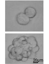
Glioma stem cells (GSC) grown as neurospheres exhibit similar characteristics to neural stem cells (NSC) grown as neurospheres, including the ability to self-renew and differentiate. GSCs are thought to play a role in cancer initiation and progression. Self-renewal potential of GSCs is thought to reflect many characteristics associated with malignancy, including tumor recurrence following cytotoxic therapy due to their proliferative dormancy and capacity to allow for the development of resistant tumor cell sub-clones due to mutations acquired during their differentiation. Here, we demonstrate that using extreme limiting dilution analysis (ELDA), subtle differences in the frequency of sphere-forming potential between PI3K-mutant oncogenic NSCs and non-oncogenic NSCs can be measured, in vitro. We further show how ELDA can be used on cells, before and after forced differentiation to amplify inherent differences in sphere-forming potential between mutant and control NSCs. Ultimately, ELDA exploits a difference in the ability of a single or a few seeded stem cells to self-renew, divide and form neurospheres. Importantly, the assay also allows a comparison between genetically distinct cells or between the same cells under different conditions, where the impact of target-specific drugs or other novel cancer stem cell therapies can be tested.
Keywords: Stem cells (干细胞)Background
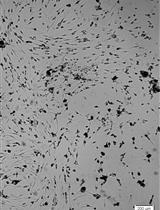
Glioblastoma (GBM) is one of the most common types of brain cancer with an extremely poor prognosis (Kaye and Morokoff, 2014). GBM treatment success or failure is determined by a complex biology due to the underlying genetics and epigenetics which regulate many signal transduction pathways, which in turn govern pro-oncogenic tumor cell behaviors and ultimately response/resistance to treatments. One of the major pathways regulating GBM cell behavior involves hyperactivation of PI3K signaling due mutations involving one or more pathway factors including the epidermal growth factor receptor (EGFR), the PI3K catalytic subunit encoded by the PIK3CA gene and deletion of the PI3K pathway negative regulator and tumor suppressor, Phosphatase and Tensin Homolog (PTEN) (Mantamadiotis, 2017). Using a genetically engineered mouse model, our recent study demonstrated that PI3K mutations, specifically targeting the Pik3ca and Pten genes in neural stem/progenitor cells (NSPs) can trigger the development of aggressive brain tumors, in vivo (Daniel et al., 2017 and 2018). In vitro analysis of NSCs isolated from mutant and wild-type mice revealed intrinsic cellular differences between genotypes, including sphere forming potential, which is a correlate of stem cell potential. This protocol describes the method used to investigate neural stem cell potential via established and variations on established methods. Extreme limiting dilution analysis (ELDA) can be used to analyze stem cell self-renewal ability and has been widely used in the stem cell field (Shackleton et al., 2006; Quintana et al., 2008; Vaillant et al., 2008). Subtle differences between NSCs or GSCs can be quantified by ELDA using a freely available webtool (Hu and Smyth, 2009). Moreover, we present a variation in stem cell potential analysis by including an intermediate differentiation step between ELDA measurements, which revealed more substantial differences in sphere forming potential between mutant and wild-type mouse NSCs, compared to performing ELDA alone, with cells which had not undergone forced differentiation. The forced differentiation step measures the inherent stability of NSCs and GBM-like NSCs, which maintain neurosphere forming potential following differentiation; stability which may correlate with faster disease relapse/recurrence following therapy.
Materials and Reagents
- Pipette tips: 10 ml, 5 ml, 2 ml (Axygen)
- 24-well tissue culture treated plate (Corning, catalog number: 3524 ) (Storage condition: 15 °C to 25 °C)
- 6-well tissue culture treated plate (Corning, catalog number: 3506 ) (Storage condition: 15 °C to 25 °C)
- 96-well Ultra-Low Attachment plate (Corning, catalog number: 3474 ) (Storage condition: 15 °C to 25 °C)
- 40 μm cell strainer (Corning, catalog number: 352340 ) (Storage condition: 15 °C to 25 °C)
- PIK3CAH1047R; PTENlox/lox; UBC-CreERT2 mice (Daniel et al., 2017 and 2018), aged E13.5 or 6 weeks old
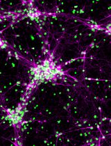
Note: NSPCs can be isolated more efficiently from younger mice. Mice used here were on a C57BL/6 genetic background. - (Optional) Antibody
- anti-GFAP(glial fibrillary protein)
- anit-Tuj1
- DMEM F12 (Thermo Fisher Scientific, GibcoTM, catalog number: 10565042 ) (storage condition: 4 °C)
- Accutase cell detachment solution (BD, catalog number: 561527 ) (storage condition: 4 °C)
- Fetal Calf serum (FCS) (Bovogen Biologicals, catalog number: SFBS-FR ) (storage condition: -20 °C)
- Penicillin-streptomycin (10,000 U/ml) (Thermo Fisher Scientific, GibcoTM, catalog number: 15140-122 ) (storage condition: -20 °C)
- B-27 supplement (50x) (Thermo Fisher Scientific, GibcoTM, catalog number: 17504-044 ) (storage condition: -20 °C)
- Epidermal growth factor (EGF) (Thermo Fisher Scientific, GibcoTM, catalog number: PHG0313 ) (storage condition: -20 °C)
- Basic fibroblast growth factor (bFGF) (BioVision, catalog number: 4036 ) (storage condition: -20 °C)
- Laminin (Thermo Fisher Scientific, GibcoTM, catalog number: 23017-015 ) (storage condition: -20 °C)
- NaCl (Sodium Chloride, Thermo Fisher Scientific, Ajax Finechem, Univar, catalog number: AJA465-500G )
- KCl (Postassium Chloride, Merck, catalog number: 1049360500 )
- Na2HPO4 (di-Sodium hydrogen phosphate, Merck, catalog number: 106586 )
- KH2PO4 (Potassium dihydrogen orthophosphate, Thermo Fisher Scientific, Ajax Chemicals, catalog number: 392 )
- Phosphate buffered saline (PBS, see Recipes)
- Neurosphere culture media (see Recipes)
- Differentiated media (see Recipes)
Equipment
- Pipettes (METTLER TOLEDO, Pipet-X, model: PX-100 )
- Biohazard safety cabinet (EuroClone, model: BioAir-TopSafe 1.2-ABC )
- Centrifuge (Dynamica Scientific, model: Velocity 18R )
- DigiRetina16 camera (Tucsen)
- Neubauer Hemocytometer (Sigma-Aldrich, catalog number: Z359629 )
- Water bath at 37 °C (Froilabo, model: BMUTE)
- Humidified incubator at 37 °C, 5% CO2 (Thermo Fisher Scientific, Heraeus, model: HeracellTM 150 )
- Inverted phase contrast microscope (Nikon, model: Diaphot , with ELWD 0.3 phase contrast)
Software
- ELDA online software (http://bioinf.wehi.edu.au/software/elda/)
Procedure
文章信息
版权信息
© 2018 The Authors; exclusive licensee Bio-protocol LLC.
如何引用
Nguyen, H. P., Daniel, P. M., Filiz, G. and Mantamadiotis, T. (2018). Investigating Neural Stem Cell and Glioma Stem Cell Self-renewal Potential Using Extreme Limiting Dilution Analysis (ELDA). Bio-protocol 8(17): e2991. DOI: 10.21769/BioProtoc.2991.
分类
干细胞 > 成体干细胞 > 神经干细胞
癌症生物学 > 癌症干细胞 > 细胞生物学试验 > 自我更新
细胞生物学 > 基于细胞的分析方法 > 非贴壁培养
您对这篇实验方法有问题吗?
在此处发布您的问题,我们将邀请本文作者来回答。同时,我们会将您的问题发布到Bio-protocol Exchange,以便寻求社区成员的帮助。