

Preparation of Cerebellum Granule Neurons from Mouse or Rat Pups and Evaluation of Clostridial Neurotoxin Activity and Their Inhibitors by Western Blot and Immunohistochemistry
用老鼠或鼠仔制备小脑颗粒神经元并通过免疫印迹和免疫组织化学法评估梭菌神经毒素活性及其抑制剂


发布: 2018年07月05日第8卷第13期 DOI: 10.21769/BioProtoc.2918 浏览次数: 11788
评审: Andrea PuharXuecai GeAnonymous reviewer(s)
参见作者原研究论文
The authors used this protocol in:
Sep 2014


Abstract
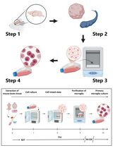
Cerebellar Granule Neurons (CGN) from post-natal rodents have been widely used as a model to study neuronal development, physiology and pathology. CGN cultured in vitro maintain the same features displayed in vivo by mature cerebellar granule cells, including the development of a dense neuritic network, neuronal activity, neurotransmitter release and the expression of neuronal protein markers. Moreover, CGN represent a convenient model for the study of Clostridial Neurotoxins (CNT), most notably known as Tetanus and Botulinum neurotoxins, as they abundantly express both CNT receptors and intraneuronal substrates, i.e., Soluble N-ethylmaleimide-sensitive factor activating protein receptors (SNARE proteins). Here, we describe a protocol for obtaining a highly pure culture of CGN from postnatal rats/mice and an easy procedure for their intoxication with CNT. We also illustrate handy methods to evaluate CNT activity and their inhibition.
Keywords: Cerebellar granule neurons (小脑颗粒神经元)Background
The large family of Clostridial Neurotoxins (CNT) is formed by Tetanus Neurotoxin (TeNT) and the many variants of Botulinum Neurotoxins (BoNT) which are the neuroparalytic toxins responsible for tetanus and botulism, respectively (Schiavo et al., 2000; Johnson and Montecucco, 2008; Rossetto et al., 2014). TeNT, the seven BoNT serotypes (BoNT/A to /G) and their many subtypes are metalloproteases that cause neuroparalysis by blocking neurotransmitter release via the cleavage of SNARE proteins (Soluble N-ethylmaleimide-sensitive factor activating protein receptors), the three essential proteins governing the fusion of synaptic vesicle with the presynaptic plasma membrane (Rossetto et al., 2014; Montecucco and Rasotto, 2015; Pirazzini et al., 2017). In addition, some putative novel serotypes (BoNT/X and BoNT/En aka eBoNT/J) (Zhang et al., 2017; Brunt et al., 2018; Zhang et al., 2018) and a BoNT-like toxin (BoNT/Wo) (Zornetta et al., 2016) displaying metalloprotease activity against SNARE proteins have been recently identified (Azarnia Tehran and Pirazzini, 2018). Yet, whether they are naturally produced and can be considered true BoNT still requires validation. Each toxin has a selective action against one specific protein that is cleaved at a distinct peptide bond (Binz, 2013; Pantano and Montecucco, 2014; Zornetta et al., 2016; Zhang et al., 2017; Zhang et al., 2018): BoNT/B, /D, /F /G, /Wo and /X hydrolyze VAMP-1/2 (vesicle-associated membrane protein) while BoNT/A and BoNT/E cleave the membrane protein SNAP-25 (synaptosomal-associated protein of 25 kDa). BoNT/C and BoNT/En are unique as they cleave more than one SNARE type: BoNT/C cleaves SNAP-25 and many isoforms of syntaxin (Pirazzini et al., 2017; Zanetti et al., 2017); instead BoNT/En cleaves different members of VAMP family and SNAP-25/23 (Zhang et al., 2018). To reach their intraneuronal substrates, CNT carry out a very sophisticated mechanism of intoxication aimed at delivering the catalytic part of the toxin within the cytosol of nerve terminals (reviewed in [Montal, 2010; Rossetto et al., 2014; Pirazzini et al., 2017]).
This process relies on cardinal functions of neuron physiology which are exploited by BoNT to enter the neuron: the expression of appropriate glycolipid and protein receptors for binding (Binz and Rummel, 2009; Rummel, 2017), the recycling of synaptic vesicles for internalization (Matteoli et al., 1996; Harper et al., 2011; Colasante et al., 2013), the generation of an electrochemical gradient across synaptic vesicle membrane to translocate the catalytic domain in the cytosol (Montal, 2010; Pirazzini et al., 2016) and the presence of a redox-chaperone system to enable SNARE proteins’ cleavage (Pirazzini et al., 2018). These features are fully preserved by cultured Cerebellar Granule Neurons (CGN), a primary culture of cerebellar granule cells from post-natal rodent cerebellum. The cerebellar cortex is composed of a few neuronal types like Purkinje cells, inhibitory interneurons and granule cells that form a highly organized tissue with well-characterized neuronal circuitries (Bilimoria and Bonni, 2008). Cerebellar granule cells constitute the most numerous and homogeneous neuronal population and can be easily isolated (Messer, 1977). Cultured CGN recapitulate many characteristics of development and maturation observed in vivo and have been extensively used as a useful model to study basic molecular and biological processes of neuron physiology like apoptosis, migration and differentiation (Contestabile, 2002).
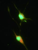
Many neuronal models, including spinal cord neurons, hiPSC derived neurons, mES derived neurons, hippocampal neurons, cortical neurons and several methods have been developed to study BoNT activity in vitro (Pellett, 2013).In our laboratory, we choose CGN as their preparation is relatively simple, rapid and very reliable and it provides a highly pure (more than 95%) and homogeneous (mostly granule cells) neuronal culture model to conveniently evaluate CNT activity by monitoring the cleavage of SNARE proteins via Western blotting or immunocytochemistry (Pirazzini et al., 2011; Eleopra et al., 2013; Pirazzini et al., 2013b and 2013c). Moreover, CGN can be adapted to the investigation of putative inhibitors and can be used as a solid platform for screening anti-BoNT antitoxins (Pirazzini et al., 2013a and 2014; Azarnia Tehran, et al., 2015; Zanetti et al., 2015; Pirazzini and Rossetto, 2017). Here, we describe a simple protocol for fast isolation of CGN, an easy procedure for their intoxication with CNT, and two methods (Western blot and immunocytochemistry) to evaluate their toxicity and inhibition.
Materials and Reagents
- 0.22 µm filters (33 mm – Merck, Millex, catalog number: SLGP033RS )
- Culture plates
6 wells (Corning, Falcon®, catalog number: 353046 )
24 wells (Corning, Falcon®, catalog number: 353047 )
96 wells (Corning, Falcon®, catalog number: 353072 ) - 35 mm Petri dishes (Corning, Falcon®, catalog number: 353001 )
- 50 ml sterile conical plastic tubes (Corning, Falcon®, catalog number: 352070 )
- Coverslips 13 mm #1 (Thermo Fisher Scientific, catalog number: 1014355113NR1 )
- Micropipette tips
1,000 µl tips (SARSTEDT, catalog number: 70.762.211 )
200 µl tips (SARSTEDT, catalog number: 70.760.211 )
10 µl tips (SARSTEDT, catalog number: 70.1130.210 ) - Nitrocellulose membranes (Sartorius, Stedim Biotech, catalog number: 11306-41BL )
- Plastic Pasteur pipettes (LP ITALIANA, catalog number: 133030 )
- Serological plastic pipettes:
5 ml plastic pipettes (Corning, Falcon®, catalog number: 357543 )
10 ml plastic pipettes (Corning, Falcon®, catalog number: 357551 )
25 ml plastic pipettes (Corning, Falcon®, catalog number: 357525 ) - Single-use stericups 0.22 µm filters ExpressTM Plus (Merck, catalog number: SCGPU0SRE )
- Sterile scalpel (CHEMIL, catalog number: 11230028 )
- Mice or rats (any gender) postnatal day 4-6
- Distilled H2O
- Ethanol (Sigma-Aldrich, catalog number: 46139 )
- Poly-L-lysine hydrobromide solution (Sigma-Aldrich, catalog number: P8920 )
- Phenol red (Sigma-Aldrich, catalog number: P3532 )
- Potassium chloride (KCl) (Sigma-Aldrich, catalog number: P5405 )
- Sodium chloride (NaCl) (Sigma-Aldrich, catalog number: S3014 )
- D-(+)-Glucose (Sigma-Aldrich, catalog number: G7528 )
- Sodium phosphate monobasic monohydrate (NaH2PO4) (Sigma-Aldrich, catalog number: S3522 )
- KH2PO4 (Sigma-Aldrich, catalog number: P5655 )
- HEPES (Sigma-Aldrich, catalog number: H3375 )
- Calcium chloride dihydrate (CaCl2·2H2O) (Sigma-Aldrich, catalog number: C3306 )
- Magnesium sulfate heptahydrate (MgSO4·7H2O) (Sigma-Aldrich, catalog number: 63138 )
- Fatty acid free bovine serum albumin (Sigma-Aldrich, catalog number: A7030 )
- Trypsin from porcine pancreas (Sigma-Aldrich, catalog number: T4799 )
- Trypsin inhibitor from soybean (Sigma-Aldrich, catalog number: T9003 )
- CompleteTM Mini, EDTA-free Protease Inhibitor Cocktail (Sigma-Aldrich, Roche Diagnostics, catalog number: 04693159001 )
- Trypan blue solution, 0.4% (Thermo Fisher Scientific, catalog number: 15250061 )
- Deoxyribonuclease I from bovine pancreas (Sigma-Aldrich, catalog number: D5025 )
- Basal Medium Eagle (BME) (Thermo Fischer Scientific, GibcoTM, catalog number: 21010046 )
- GlutamaxTM Supplement (Thermo Fischer Scientific, GibcoTM, catalog number: 35050061 )
- Gentamicin solution (Sigma-Aldrich, catalog number: G1272 )
- Cytosine β-D-arabinofuranoside (AraC) (Sigma-Aldrich, catalog number: C1768 )
- Fetal Bovine Serum (FBS) (EUROCLONE, catalog number: EUS 028877 )
Note: Comparative testing for culture optimization is needed when changing serum lot or supplier. - Trizma® base (Sigma-Aldrich, catalog number: T1503 )
- Trizma® hydrochloride (Sigma-Aldrich, catalog number: T3253 )
- Glycine (Sigma-Aldrich, catalog number: G8898 )
- Sodium dodecyl sulfate (SDS, Sigma-Aldrich, catalog number: L3771 )
- Bromophenol Blue (Sigma-Aldrich, catalog number: B0126 )
- Glycerol (Sigma-Aldrich, catalog number: G9012 )
- 2-mercaptoethanol (Sigma-Aldrich, catalog number: M6250 )
- NuPAGETM 12% Bis-Tris Gels (Thermo Fisher Scientific, catalog number: NP0341BOX )
- NuPAGETM 4-12% Bis-Tris Gels (Thermo Fisher Scientific, catalog number: NP0321BOX )
- NuPAGETM MES SDS Running Buffer (20x) (Thermo Fisher Scientific, catalog number: NP0002 )
- NuPAGETM MOPS SDS Running Buffer (20x) (Thermo Fisher Scientific, catalog number: NP0001 )
- Methanol (Sigma-Aldrich, catalog number: 322415 )
- Ponceau S (Sigma-Aldrich, catalog number: P3504 )
- Acetic acid (Sigma-Aldrich, catalog number: 45726 )
- Tween 20® (Sigma-Aldrich, catalog number: P9416 )
- Anti Syntaxin-1A/1B antibody (homemade)
- Anti-BoNT/A-cleaved SNAP-25 antibody (homemade)
- Anti-BoNT/B-TeNT-cleaved VAMP2 antibody (homemade)
- Anti-BoNT/E-cleaved SNAP-25 antibody (homemade)
- Anti-SNAP-25 antibody (SMI81, Abcam, catalog number: ab24737 )
- Anti-Syntaxin1 antibody (Synaptic System, catalog number: 110 011 )
- Anti-VAMP-2 antibody (Synaptic System, catalog number: 104 211 )
- Paraformaldehyde (Sigma-Aldrich, catalog number: P6148 )
- Ammonium chloride (Sigma-Aldrich, catalog number: A9434 )
- Fluorescence mounting medium (Agilent Technologies, Dako, catalog number: S3023 )
- HRP-conjugated or fluorescent-conjugated secondary antibodies (any supplier)
- Sodium dodecyl sulfate (Sigma-Aldrich, catalog number: 74255 )
- Tetanus neurotoxin and botulinum neurotoxins. The neurotoxins used in our laboratory are purified as previously described (Schiavo and Montecucco, 1995; Shone and Tranter, 1995) or produced recombinantly (Bade et al., 2004; Zanetti et al., 2017)
- Krebs solution (10x) (see Recipes)
- 155 mM MgSO4 stock solution (see Recipes)
- 12.2 mM CaCl2 stock solution (see Recipes)
- Solution A (see Recipes)
- Solution B (see Recipes)
- Solution C (see Recipes)
- Solution D (see Recipes)
- Solution E (see Recipes)
- Poly-L-lysine solution (see Recipes)
- BME complete medium, CGN culture medium (see Recipes)
- Cytosine β-D-arabinofuranoside (AraC) stock solution (see Recipes)
- PBS (see Recipes)
- PBST (see Recipes)
- SDS-PAGE Sample loading buffer (4x) (see Recipes)
- Transfer buffer (see Recipes)
- Ponceau S solution (see Recipes)
- Blocking buffer (see Recipes)
- 4% Paraformaldehyde solution (see Recipes)
- Quenching solution (see Recipes)
- Permeabilization solution (see Recipes)
- Blocking solution (see Recipes)
Equipment
- Micropipettes (any supplier)
- Pipette controller (any supplier)
- Centrifuge for conical tubes (Eppendorf, model: 5804 R )
- Dissecting hood (any supplier)
- Dissecting stereomicroscope (OPTIKA Microscopes, model: SZM-LED2 )
- Sterile laminar flow hood (any supplier)
- Hemacytometer (any supplier)
- Humidified incubator (5% CO2 at 37 °C) (any supplier)
- Ice bucket and ice
- Scissors #1 (Rudolf Medical GmbH, catalog number: RU 2675-18 )
- Forceps #2 (Rudolf Medical GmbH, catalog number: RU 7584-16 )
- Scissors #3 (Rudolf Medical GmbH, catalog number: RU 2246-09 )
- Tweezers #4 (Rudolf Medical GmbH, catalog number: RU 4240-05 )
- Mini gel tank (Thermo Fisher Scientific catalog number: A25977 )
- Mini Trans-Blot® Cell (Bio-Rad Laboratories, catalog number: 1703930 )
- Thermoblock (any supplier)
- Thermosettable water-bath (37 °C) (any supplier)
- Vortex mixer (any supplier)
- Epifluorescence or confocal microscope: Leica CTR6000 equipped with X5 N PL AN (Leica Microsystems, model: Leica CTR6000 )
- 0.12, x20 N PL AN 0.40 objectives or Leica SP5 equipped with 100x HCX PL APO NA 1.4 objective, respectively (Leica Microsystems, Wetzlar, Germany)
Procedure
文章信息
版权信息
© 2018 The Authors; exclusive licensee Bio-protocol LLC.
如何引用
Azarnia Tehran, D. and Pirazzini, M. (2018). Preparation of Cerebellum Granule Neurons from Mouse or Rat Pups and Evaluation of Clostridial Neurotoxin Activity and Their Inhibitors by Western Blot and Immunohistochemistry. Bio-protocol 8(13): e2918. DOI: 10.21769/BioProtoc.2918.
分类
神经科学 > 细胞机理 > 细胞分离和培养
细胞生物学 > 细胞分离和培养 > 细胞分离
生物化学 > 蛋白质 > 电泳
您对这篇实验方法有问题吗?
在此处发布您的问题,我们将邀请本文作者来回答。同时,我们会将您的问题发布到Bio-protocol Exchange,以便寻求社区成员的帮助。