

Preserve Cultured Cell Cytonemes through a Modified Electron Microscopy Fixation
通过一种改良的电子显微技术固定方法保存培养的细胞导管

发布: 2018年07月05日第8卷第13期 DOI: 10.21769/BioProtoc.2898 浏览次数: 6581
评审: Zinan ZhouTomas AparicioSilvia Caggia
参见作者原研究论文
The authors used this protocol in:
Oct 2017


Abstract
Immunocytochemistry of cultured cells is a common and effective technique for determining compositions and localizations of proteins within cellular structures. However, traditional cultured cell fixation and staining protocols are not effective in preserving cultured cell cytonemes, long specialized filopodia that are dedicated to morphogen transport. As a result, limited mechanistic interrogation has been performed to assess their regulation. We developed a fixation protocol for cultured cells that preserves cytonemes, which allows for immunofluorescent analysis of endogenous and over-expressed proteins localizing to the delicate cellular structures.
Keywords: MEM-fix (MEM固定)Background
Cytonemes are classified as thin (~200 nm diameter) actin based filopodia, over 2 μm in length, which can transport morphogens (Ramírez-Weber and Kornberg, 1999). These signaling structures were first classified and described in detail in the developing Drosophila wing imaginal disc, and have subsequently been observed in mouse, chick and zebrafish model organisms (Ramírez-Weber and Kornberg, 1999; Sanders et al., 2013; Stanganello et al., 2015). In most of these cases, cytoneme detection was only possible with live imaging of over-expressed, fluorescently labeled proteins. Examination of cytonemes of cultured cells has been limited due to traditional fixation protocols failing to preserve these fragile filaments. These complications have been limiting factors in determining the cellular mechanisms driving cytoneme formation and function during development and tissue homeostasis, and determining whether these processes are corrupted in disease.
In order to overcome these limitations, we developed a modified electron microscopy fixative (MEM-fix)-based protocol that preserves cytonemes of cultured cells. Use of MEM-fix allows for the detection of endogenous and over-expressed proteins of interest in the filopodial structures via traditional immunofluorescent protocols (Bodeen et al., 2017). MEM-fix is generated by the addition of glutaraldehyde to a final working concentration of 0.5% to a standard 4% paraformaldehyde fixative solution. Glutaraldehyde, which is commonly used to fix cells for electron microscopy-based studies, was included because of its ability to effectively preserve subcellular structures. Although glutaraldehyde is not an optimal fixative for immunofluorescence microscopy due to its propensity to auto-fluoresce, we determined that addition of 26.4 mM sodium borohydride to the permeabilization buffer was sufficient to mitigate this undesirable side effect (Tagliaferro et al., 1997; Bacallao et al., 2006). Unfortunately, due to glutaraldehyde limiting antibody penetration into cells, MEM-fix is not conducive to staining of cytoplasmic or nuclear proteins, so should be limited to an examination of integral membrane or juxta-membrane proteins.
We recently used this technique to examine Hedgehog (Hh) morphogen transport through cytonemes of cultured Drosophila cells and mouse fibroblasts (Bodeen et al., 2017). Here, we provide an optimized protocol for imaging of cytonemes in NIH3T3 cells, and provide examples of its adaptability to additional cultured mammalian cell lines (Figure 1). MEM-fix protocol modifications to standard cell fixation methods allow for reproducible detection of cytonemes and immunofluorescence-based staining of trans-membrane and membrane-adjacent proteins in cultured cells. MEM-fix also preserves signal of fluorescently-labeled proteins, so is not solely dependent on immuno-detection based methods.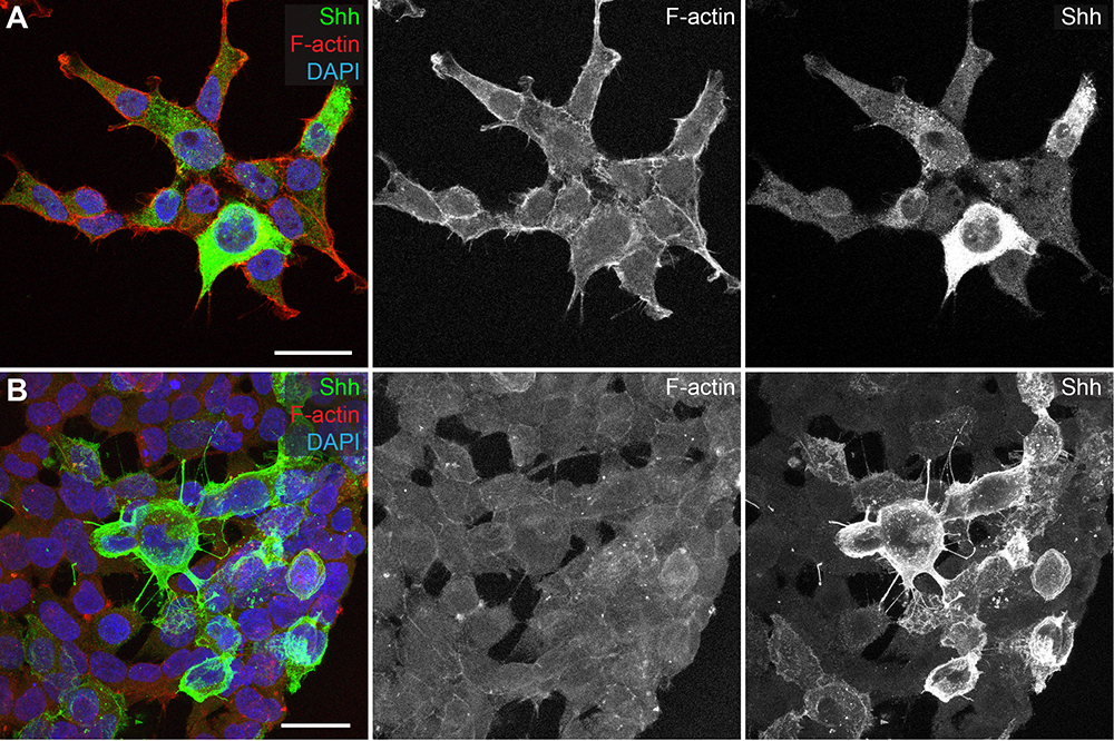
Figure 1. MEM-fix increases preservation of cytonemes in cultured cells compared to paraformaldehyde fixation. A and B. HEK293T cells transfected with Shh. A. Cells fixed with paraformaldehyde show some actin-based protrusions, but do not show Shh (green) positive filaments. B. Cells fixed with MEM-fix contain many cytoneme projections, marked by the presence of F-actin (red) and Shh. Scale bars = 25 µm.
Materials and Reagents
- SHARP® Precision Barrier Tips, For P-1000 and Eppendorf 1,000, 1,250 µl (Denville Scientific, catalog number: P1126 )
- SHARP® Precision Barrier Tips, For P-200, 200 µl (Denville Scientific, catalog number: P1122 )
- SHARP® Precision Barrier Tips, For P-20, 20µl (Denville Scientific, catalog number: P1121 )
- SHARP® Precision Barrier Tips, Extra Long for P-2 and P-10, 10 µl (Denville Scientific, catalog number: P1096-FR )
- Gold SealTM Rite-OnTM Micro Slides (Thermo Fisher Scientific, catalog number: 3050-002 )
- 12 mm Microscope Cover Glass-1.5 (Fisher Scientific, catalog number: 12-545-81 )
- TPP® centrifuge tubes, volume 50 ml, polypropylene (TPP Techno Plastic Products, catalog number: 91050 )
- TPP® centrifuge tubes, volume 15 ml, polypropylene (TPP Techno Plastic Products, catalog number: 91015 )
- StericupTM Sterile Vacuum Filter Units 500 ml (Merck, catalog number: SCGPU05RE )
- 24-well plate (Corning, Falcon®, catalog number: 353226 )
- 6-well plate NunclonTM Delta Surface (Thermo Fisher Scientific, catalog number: 140675 )
- Professional Kimtech ScienceTM KimwipesTM (KCWW, Kimberly-Clark, catalog number: 34155 )
- Premium Microcentrifuge Tubes: 1.5 ml (Fisher Scientific, catalog number: 05-408-129 )
- NIH3T3 (ATCC, catalog number: CRL-1658 )
- Ultrapure water
- DMEM (1x) 4.5 g/L D-glucose, [-] L-Glutamine, [-] HEPES, [-] Sodium Pyruvate (Thermo Fisher Scientific, GibcoTM, catalog number: 11960044 )
- Opti-MEMTM Reduced Serum Medium (Thermo Fisher Scientific, catalog number: 31985070 )
- Pen/Strep, 100x (Merck, catalog number: TMS-AB2-C )
- HyCloneTM Cosmic CalfTM Serum (BCS) (GE Healthcare, catalog number: SH30087.03 )
- MEM NEAA (100x) MEM Non-Essential Amino Acids (Thermo Fisher Scientific, GibcoTM, catalog number: 11140050 )
- L-Glutamine (100x) (100 ml) (Thermo Fisher Scientific, InvitrogenTM, catalog number: 25030081 )
- Sodium Pyruvate (100 mM) 100x (Thermo Fisher Scientific, GibcoTM, catalog number: 11360070 )
- Lipofectamine® 3000 transfection kit (Thermo Fisher Scientific, InvitrogenTM, catalog number: L3000-015 )
- 70% (volume) ethanol diluted in water
- 0.05% Trypsin 0.53 nM EDTA, 1x [-] Sodium Bicarbonate (Corning, catalog number: 25-052-Cl )
- DPBS, 1x (Dulbecco's Phosphate-Buffered Saline) (Corning, CellgroTM, catalog number: 21-031-CM )
- Sodium phosphate dibasic (Sigma-Aldrich, catalog number: S0876 )
- Sodium phosphate monobasic (Sigma-Aldrich, catalog number: S5011 )
- Sodium borohydride (Sigma-Aldrich, catalog number: 213462-25G )
- Formaldehyde, 16%, methanol free, Ultra Pure EM Grade (Polysciences, catalog number: 18814-10 )
- Glutaraldehyde, 8% Aqueous Solution, EM Grade (Electron Microscopy Sciences, catalog number: 16019 )
- Normal Goat Serum (10 ml) (Jackson ImmunoResearch, catalog number: 005-000-121 )
- Triton X-100 (Sigma-Aldrich, catalog number: T9284 )
- Tween 20 (Acros Organics, catalog number: AC233360010 )
- Sonic hedgehog (Shh) antibody (H-160), rabbit polyclonal (Santa Cruz Biotechnology, catalog number: sc-9024 )
- Alexa FluorTM 633 Phalloidin (Thermo Fisher Scientific, catalog number: A22284 )
- Goat anti-Rabbit IgG (H+L) Secondary Antibody, Alexa Fluor 488 (Thermo Fisher Scientific, Invitrogen, catalog number: R37116 )
- DAPI Solution (1 mg/ml) (Thermo Fisher Scientific, catalog number: 62248 )
- Prolong® Diamond Antifade Mountant (Thermo Fisher Scientific, Invitrogen, catalog number: P36961 )
- NIH3T3 media (see Recipes)
- NIH3T3 serum/antibiotic-free media (see Recipes)
- 0.2 M Dibasic Sodium Phosphate solution (see Recipes)
- 0.2 M Monobasic Sodium Phosphate solution (see Recipes)
- Modified electron microscopy fixative (MEM-fix) (see Recipes)
- Permeabilization buffer (see Recipes)
- PBGT (see Recipes)
- Primary antibody solution (see Recipes)
- Secondary antibody solution (see Recipes)
Equipment
- Eppendorf Research Plus single channel pipette, 100-1,000 µl (Eppendorf, catalog number: 3123000063 )
- Eppendorf Research Plus single channel pipette, 20-200 µl (Eppendorf, catalog number: 3123000055 )
- Eppendorf Research Plus single channel pipette, 0.5-10 µl (Eppendorf, catalog number: 3123000020 )
- Eppendorf Research Plus single channel pipette, 0.1-2.5 µl (Eppendorf, catalog number: 3123000012 )
- Allegra X-12R centrifuge (Beckman Coulter, model: Allegra® X-12R , catalog number: 392302)
- Heracell VIOS 160i CO2 incubator (at 37 °C and 5% CO2), (Thermo Fisher Scientific, model: HeracellTM VIOS 160i, catalog number: 51030287 )
- AE50 analytical balance (Mettler-Toledo International, model: AE50 )
- Aspirator
- Precision dual-chamber water bath 288 (Thermo Fisher Scientific, catalog number: 2853 )
- Biological safety cabinet (The Baker Company, catalog number: B40-112 )
- Cell counting chamber (Hausser Scientific, catalog number: 3200 )
- FisherbrandTM Fine Point High Precision Forceps (Fisher Scientific, catalog number: 22-327379 )
- Confocal laser-scanning microscope (Leica Microsystems, model: Leica TCS SP8 )
Software
- Leica Application Suite X (used to generate Tiffs)
Procedure
文章信息
版权信息
© 2018 The Authors; exclusive licensee Bio-protocol LLC.
如何引用
Hall, E. T. and Ogden, S. K. (2018). Preserve Cultured Cell Cytonemes through a Modified Electron Microscopy Fixation. Bio-protocol 8(13): e2898. DOI: 10.21769/BioProtoc.2898.
分类
发育生物学 > 细胞信号传导 > 命运决定
发育生物学 > 形态建成 > 器官形成
细胞生物学 > 细胞成像 > 固定细胞成像
您对这篇实验方法有问题吗?
在此处发布您的问题,我们将邀请本文作者来回答。同时,我们会将您的问题发布到Bio-protocol Exchange,以便寻求社区成员的帮助。