

Quantification of Extracellular Double-stranded RNA Uptake and Subcellular Localization Using Flow Cytometry and Confocal Microscopy
流式细胞术和共聚焦显微镜检查定量测定细胞外双链RNA摄取和亚细胞定位
发布: 2018年06月20日第8卷第12期 DOI: 10.21769/BioProtoc.2890 浏览次数: 7625
评审: Ivan ZanoniLaura CampisiKristofor K. Ellestad
参见作者原研究论文
The authors used this protocol in:

Sep 2017


Abstract
Double-stranded RNA is a potent pathogen-associated molecular pattern (PAMP) produced as a by-product of viral replication and a well-known hallmark of viral infection. Viral dsRNAs can be released from infected cells into the extracellular space and internalized by neighboring cells via endocytosis. Mammals possess multiple pattern recognition receptors (PRRs) capable of detecting viral dsRNAs such as endosomal toll-like receptor 3 (TLR3) and cytosolic RIG-I-like receptors (RLRs) which lead to the production of type I interferons (IFNs). Thus, intracellular localization of viral dsRNA can provide insight into the downstream signaling pathways leading to innate immune activation. Here, we describe a quantitative method for measuring extracellular dsRNA uptake and visualizing subcellular localization of internalized dsRNA via flow cytometry and confocal microscopy respectively.
Keywords: Double-stranded RNA (双链RNA)Background
Double-stranded RNAs (dsRNAs) are a common by-product of viral replication and are potent activators of antiviral immunity via the production of type I interferon (IFN) and other pro-inflammatory cytokines (Nellimarla and Mossman, 2014). Viral dsRNAs are sensed within endosomes by TLR3 (Matsumoto et al., 2003) or in the cytosol by the RIG-I-like receptors (RLRs), RIG-I and MDA-5 (Kato et al., 2006). During lytic infections, these dsRNAs can be released into the extracellular space where they bind surface receptors on neighboring cells, such as class A scavenger receptors (SR-A) and Raftlin, and are subsequently internalized via clathrin-mediated endocytosis (Itoh et al., 2008; DeWitte-Orr et al., 2010; Watanabe et al., 2011; Dansako et al., 2013).
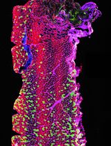
In our previous study, we found out that the protein SID1 transmembrane family member 2 (SIDT2) localizes to late endosomes and lysosomes and that loss of SIDT2 leads to subcellular accumulation of the synthetic dsRNA analog, poly(I:C), while not affecting initial endocytosis-mediated internalization (Nguyen et al., 2017). To do so, we developed and utilized flow cytometry and confocal microscopy-based approaches to quantitatively measure poly(I:C) uptake and subcellular localization respectively in vitro. In this protocol, we describe a further refinement of these assays to allow for high-throughput assessment of internalization and subcellular localization of different dsRNAs. These methods allow for further dissection of dsRNA trafficking during viral infection and the downstream effects of these dsRNAs on innate immune signaling.
Materials and Reagents
- 8 well microscope slide (ibidi, catalog number: 80826 )
- 24 well tissue culture plate (Corning, Falcon®, catalog number: 353047 )
- Sterile filtered pipette tips (0.5 µl to 1,000 µl) (Corning, Axygen, catalogue numbers: TF-300-L-R-S , TF-20-L-R-S , TF-200-L-R-S and TF-1000-L-R-S )
- 10 cm culture dishes (Corning, Falcon®, catalog number: 353003 )
- 10 ml centrifuge tubes (SARSTEDT, catalog number: 62.9924.284 )
- 1.5 ml microcentrifuge tubes (Sigma-Aldrich, catalog number: EP0030120086 )
- 1.2 ml Micro Titertube (Thermo Fisher Scientific, Quality Scientific Plastics, catalog number: 845-Q )
- Serological pipettes, individually wrapped, 10 ml (Corning, Falcon®, catalog number: 356551 )
- Mammalian cell line of interest, here: mouse embryonic fibroblasts (see Note 1)
- 70% (v/v) ethanol (Chem Supply, catalog number: EA043 )
- Dulbecco’s modified Eagle medium (DMEM) or other suitable complete growth medium for culture of cell line of interest
- Fetal bovine serum (FBS) (Sigma-Aldrich, catalog number: F9423 )
- Phosphate-buffered saline (PBS) (sterile) (Thermo Fisher Scientific, GibcoTM, catalog number: 14190250 )
- Penicillin/streptomycin solution (Sigma-Aldrich, catalog number: P4333 )
- Poly(I:C)-fluorescein (InvivoGen, catalog number: tlrl-picf )
- Poly(I:C)-rhodamine (InvivoGen, catalog number: tlrl-picr )
- dsRNA specific monoclonal antibody (J2, SCICONS English and Scientific Consulting, catalog number: 10010200 )
- RNase A enzyme (Sigma-Aldrich, catalog number: R4875 )
- 1x trypsin-EDTA solution (Sigma-Aldrich, catalog number: 59430C )
- Paraformaldehyde (PFA) powder (Sigma-Aldrich, catalog number: 158127 )
- Tween 20 (Sigma-Aldrich, catalog number: P1379 )
- DAPI (4’,6-Diamidine-2’-phenylindole dihydrochloride) powder (Sigma-Aldrich, catalog number: D9542 )
- ImmersolTM Immersion Oil (Carl Zeiss, catalog number: 4449620000000 )
- Complete growth medium (see Recipes)
- 10% FBS/PBS (see Recipes)
- 4% paraformaldehyde (w/v) (see Recipes)
- Permeabilization buffer (see Recipes)
- DAPI solution (see Recipes)
Equipment
- Pipetting aid (Thermo Fisher Scientific, catalog number: 9531 )
- Micropipettes from 0.5 µl to 1 ml (Mettler-Toledo International, Rainin, model: Pipet-LiteTM XLS+ )
- Hemocytometer
- Class II biological safety cabinet/tissue culture hood
- Humidified CO2 incubator (95% air, 5% CO2, 37 °C)
- Inverted light microscope (phase contrast)
- 37 °C water bath
- Vacuum aspiration system with glass Pasteur pipettes
- Table top centrifuge equipped with a swing-out rotor for 10 ml conical tubes
- Microcentrifuge
- LSRFortessa X20 (BD, BD Biosciences, model: LSRFortessaTM X-20 ) or equivalent flow cytometer
- LSM 780 confocal laser scanning microscope (ZEISS, model: LSM 780 ) or equivalent microscope
Software
- FIJI/ImageJ
- Zeiss ZEN package
- Microsoft Excel
- FlowJo
- GraphPad Prism 7
Procedure
文章信息
版权信息
© 2018 The Authors; exclusive licensee Bio-protocol LLC.
如何引用
Nguyen, T. A., Whitehead, L. and Pang, K. C. (2018). Quantification of Extracellular Double-stranded RNA Uptake and Subcellular Localization Using Flow Cytometry and Confocal Microscopy. Bio-protocol 8(12): e2890. DOI: 10.21769/BioProtoc.2890.
分类
免疫学 > 免疫细胞成像 > 共聚焦显微镜技术
细胞生物学 > 细胞成像 > 共聚焦显微镜
分子生物学 > RNA > RNA 检测
您对这篇实验方法有问题吗?
在此处发布您的问题,我们将邀请本文作者来回答。同时,我们会将您的问题发布到Bio-protocol Exchange,以便寻求社区成员的帮助。