

In vivo Use of Dextran-based Anterograde Cortical Tracers to Assess the Integrity of the Cortical Spinal Tract
体内使用基于右旋糖酐的顺行皮质示踪剂评估皮质脊髓束的完整性
.jpg)
发布: 2018年05月20日第8卷第10期 DOI: 10.21769/BioProtoc.2862 浏览次数: 7666
评审: Oneil G. BhalalaTrevor Martin SmithEhsan Kheradpezhouh
参见作者原研究论文
The authors used this protocol in:
Nov 2017
Advertisement


Abstract
When injected into the motor cortex of rats, anterograde tracers label fibers of the associated descending corticospinal tract (CST) that originate from pyramidal neurons in the tracer-injected cortex. These fibers can be assessed at the level of the spinal cord to determine the integrity of the descending CST and the spatial distribution of axons in the spinal grey matter. Here we provide detailed methods on the minimally invasive stereotaxic injection of anterograde tracers into the forelimb sensorimotor representation in the rat cortex. In addition, we detail the fixing and processing of spinal tissue for assessment of CST integrity and branching into spinal grey matter.
Keywords: Cortical spinal tract (皮质脊髓束)Background
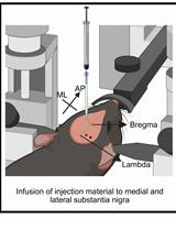
The motor cortex contains upper motor neurons which give rise to descending axons that form the CST and terminate in the spinal cord. Anterograde tracers injected into the motor cortex of rats label these descending fibers of the CST allowing their terminals to be assessed at the level of the spinal cord (Figure 1). In rodent models, anterograde labeling of the CST is widely used to assess CST integrity in a range of spinal cord injury models including lesions (Thallmair et al., 1998; Vavrek et al., 2006) and contusions (Hill et al., 2001). Further, anterograde cortical tracers have been used to assess spared fibers and plasticity of the CST in various models of brain injury such as cortical stroke (Wahl et al., 2014; Wiersma et al., 2017), traumatic brain injury (Ueno et al., 2012) and pyramidotomy (Starkey et al., 2012). Here we provide detail methods on injection of anterograde tracers into motor cortex corresponding the forelimb. We describe how to create burr holes without inducing damage to the cortex and the use of pulled glass pipette tips for minimally invasive injection. Since the CST is widely assessed in both brain and spinal cord injury models, we offer a systematic method of assessing the CST and branching fibers in the surrounding spinal grey matter that accounts for variability due to inter-animal differences in tracer efficacy or uptake.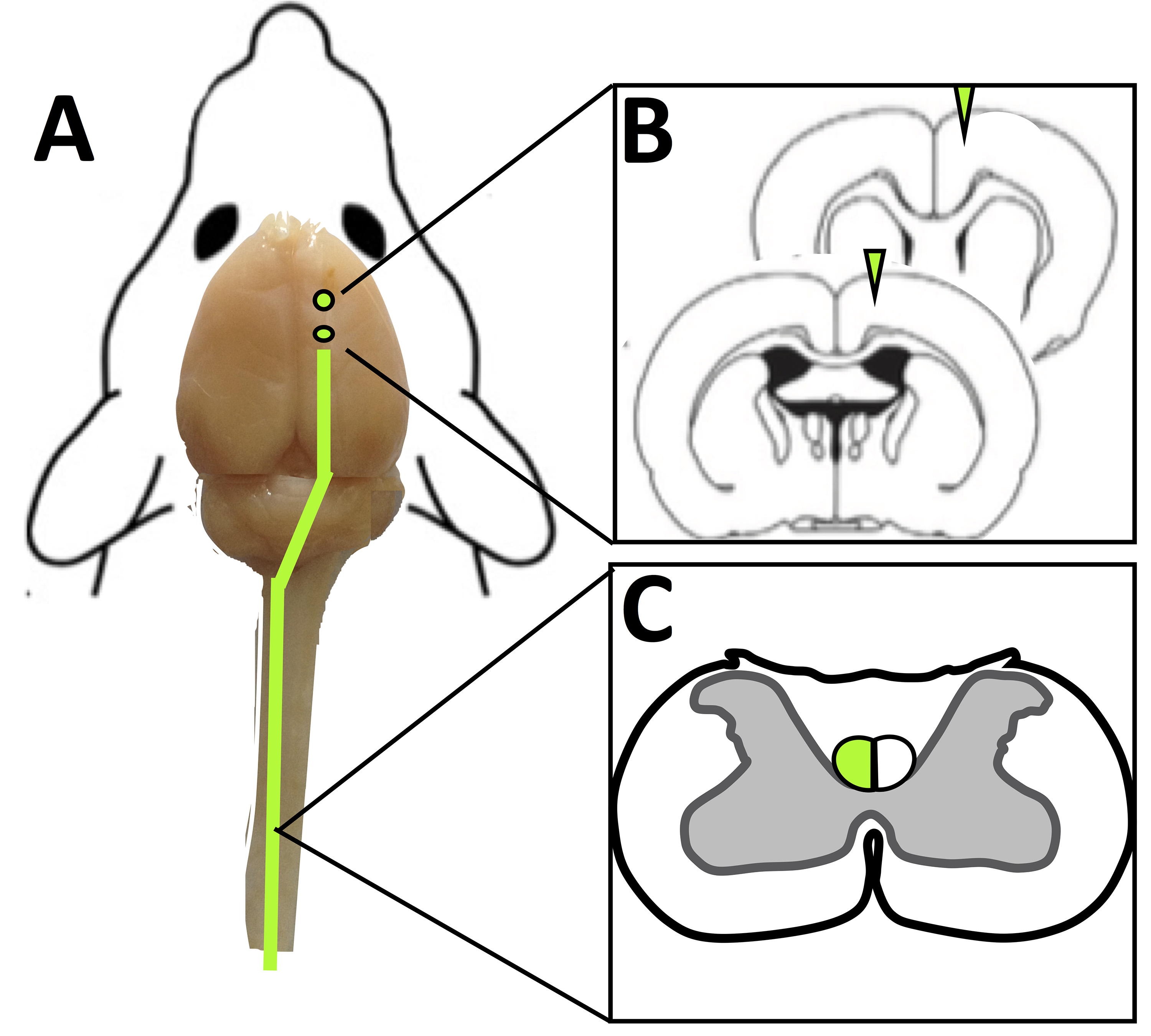
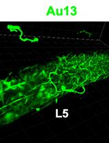
Figure 1. Use of anterograde cortical tracers to label the corticospinal tract in rats. A. Tracer injection sites in the rat cortex (shown as green circle) and anterograde tracer labeled descending fibers (green line). B. Coronal sections of the brain at tracer injection sites. C. Transverse slices of the spinal cord (C4) with tracer labeled fibers of the CST shown in green.
Materials and Reagents
- Surgical
- Sterile scalpel blade # 10 (Fine Scientific Tools, catalog number: 10100-00 )
- 10 µl Hamilton syringe (Hamilton, catalog number: 80366 )
- Glass microtubule–Glass filament with 1.0 mm external diameter (Sutter Instrument, catalog number: BF150-86-10 )
- 16-gauge needle (STEMCELL Technologies, catalog number: 28110 )
- 1 ml syringes x 5 (BD, catalog number: 309659 )
- Silk suture 5-0, P-3 reverse cutting (Ethicon, PERMAHAND®, catalog number: 640G )
- Sprague Dawley Rat, male, ~500 g,15-20 weeks of age (Charles River)
- Isoflurane USP 99% (Fresenius Kabi, catalog number: CP0406V2 )
- Compressed oxygen gas high purity 99.995% (Praxair)
- Compressed nitrous oxide gas 99% purity (Praxair)
- Sterile saline, 0.9% NaCl (Baxter, catalog number: 2B1324X )
- 0.25% Bupivacaine (Sigma-Aldrich, catalog number: B5274 )
- Buprenorphine (Schering- Plough, 0.2 mg injectable)
- Sterile scalpel blade # 10 (Fine Scientific Tools, catalog number: 10100-00 )
- Tracer
- Dextran, Alexa FluorTM 488, 10,000 MW, Anionic, Fixable (Thermo Fisher Scientific, catalog number: D22910 )
- Dextran amine, Texas Red®, 10,000 MW (Vector Laboratories, catalog number: SP-1140 )
- Dextran, Alexa FluorTM 488, 10,000 MW, Anionic, Fixable (Thermo Fisher Scientific, catalog number: D22910 )
- Perfusion
- Euthasol® (VIRBAC, catalog number: 710101 )
- Celline isotonic saline solution (Fisher Scientific, catalog number: 351142-10)
Manufacturer: SCP SCIENTIFIC, catalog number: CS20310D . - Heparin (Sigma-Aldrich, catalog number: H3393 )
- Formalin 1:10 dilution, buffered (Fisher Scientific, catalog number: SF100-20 )
- 4% formalin solution (see Recipes)
- Isotonic saline heparin (5,000 IU/L) solution (see Recipes)
- Euthasol® (VIRBAC, catalog number: 710101 )
- Tissue cryosectioning
- Superfrost® Plus Gold slides (Fisher Scientific, catalog number: 22-035813 )
- Isopentane (2-methylbutane) C5H12 (Sigma-Aldrich, catalog number: 277258 )
- D-Sucrose (Fisher Scientific, catalog number: 10638403 )
- Tissue Tek® Optimal cutting temperature (OCT) compound (Sakura, catalog number: 4583 )
- VECTASHIELD (Vector Laboratories, catalog number: H-1000 )
- 30% Sucrose solution (see Recipes)
- Superfrost® Plus Gold slides (Fisher Scientific, catalog number: 22-035813 )
- Spinal cord analysis
- Rat atlas (Paxinos, George, and Charles Watson. The rat brain in stereotaxic coordinates: hard cover edition. Access Online via Elsevier, 2006. A tool by Matt Gaidica.)
Equipment
- Heating pad (Stoelting, catalog number: 50300 )
- Betadine® surgical scrub–(Fisher Scientific, catalog number: 19-027132 )
- Sterile Crile Hemostats (Fine Science Tools, catalog number: 13004-14 )
- Sterile blunt ended scissors (Fine Science Tools, catalog number: 14001-18 )
- Sterile rongeur (Fine Science Tools, catalog number: 16000-14 )
- Carbide burs drill bit (SS WHITE, catalog number: 14002-5 )
- Perfusion tray (Thermo Fisher Scientific, Thermo ScientificTM, catalog number: 11469 )
- Sterile scoop (Fine Science Tools, catalog number: 10090-13 )
- Weigh scale (Kent Scientific, catalog number: SCL-1015 )
- Animal Anesthetic System (Harvard Apparatus, catalog number: 72-6467 )
- Stereotaxic frame (KOPF INSTRUMENTS, model: Model 963 with standard accessories)
- Micropipette puller (Sutter Instrument, catalog number: P-30 )
- Surgical drill–TCM Endo III (Nouvag, catalog number: 31817 )
- Digital Peri-Star Pro-Peristaltic Pump (World precision instruments, catalog number: PERIPRO-4HS )
- Cryostat (Leica Biosystems, catalog number: CM3050 S research cryostat)
- Leica HCX PL APO L 10x 1.0NA water emersion objective lens
- Leica SP5 Confocal microscope (Leica Microsystems, model: Leica SP5 )
- Inverted microscope (Leica Microsystems, catalog number: Leica DMI6000B )
- Animal surgery stereo microscope (Leica, M-series)
Software
- Leica LAS AF Software
- ImageJ
- Prism by Graph Pad vs9
Procedure
文章信息
版权信息
© 2018 The Authors; exclusive licensee Bio-protocol LLC.
如何引用
Readers should cite both the Bio-protocol article and the original research article where this protocol was used:
- Wiersma, A. M. and Winship, I. R. (2018). In vivo Use of Dextran-based Anterograde Cortical Tracers to Assess the Integrity of the Cortical Spinal Tract. Bio-protocol 8(10): e2862. DOI: 10.21769/BioProtoc.2862.
- Wiersma, A. M., Fouad, K. and Winship, I. R. (2017). Enhancing spinal plasticity amplifies the benefits of rehabilitative training and improves recovery from stroke. J Neurosci 37(45): 10983-10997.
分类
神经科学 > 神经系统疾病 > 动物模型
神经科学 > 神经解剖学和神经环路 > 动物模型
您对这篇实验方法有问题吗?
在此处发布您的问题,我们将邀请本文作者来回答。同时,我们会将您的问题发布到Bio-protocol Exchange,以便寻求社区成员的帮助。