

An Image-based Assay for High-throughput Analysis of Cell Proliferation and Cell Death of Adherent Cells
采用基于成像的分析法进行粘附细胞的细胞增殖和细胞死亡高通量分析


发布: 2018年05月05日第8卷第9期 DOI: 10.21769/BioProtoc.2835 浏览次数: 10994
评审: Alessandro DidonnaMichela PeregoSilvia Caggia
参见作者原研究论文
The authors used this protocol in:
Dec 2017
Advertisement


Abstract
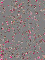
In this protocol, we describe a method to monitor cell proliferation and death by live-cell imaging of propidium iodide (PI)-stained adherent mammalian cells. PI is widely used to assess cell death. However, it is usually used in end-point assays. Recently, we implemented the use of PI for real-time cell death assessment by automated imaging. Cells are seeded in a 96-well format, and after attachment, the treatments are added directly to the wells together with PI. Thereafter, cells are subjected to automated time-lapse imaging and quantification by computer software. Combined analyses of phase-contrast and fluorescence images allow assessment of treatment effects on cell proliferation as well as the extent and kinetics of cell death.
Keywords: Cell death (细胞死亡)Background
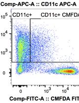
A variety of cell-based assays are available to determine cell death, but most of them, including the MTT (3-(4,5-dimethylthiazol-2-yl)-2,5-diphenyltetrazolium bromide) assay, crystal blue staining, and various flow cytometry-based methods, have the limitation of being end-point assays. Recently, we employed propidium iodide (PI) for live-cell assessment of cell death using an automated fluorescence imaging system (IncuCyte ZOOM from Essen Bioscience) (Sehgal et al., 2017). By combined analysis of phase-contrast images, cytostatic and cytotoxic effects can be simultaneously detected. This method proved to be reliable and reproducible, as well as very simple and cheap. In our recent publication, we used it in three different cancer cell lines (LNCaP, PC3 and MCF7) to efficiently determine and compare the toxicities of various drug analogs of the ER Ca2+ pump inhibitor thapsigargin (Tg) (Sehgal et al., 2017). The use of PI for real-time analysis of cell death has been described and validated by others (Wlodkowic et al., 2009; Zhao et al., 2010), but the analysis methods employed previously (e.g., flow cytometry, or lab-on-chip platforms) are more laborious and time-consuming than the simple analysis method we present here. In short, we quantify cell death using the integrated algorithms of the IncuCyte ZOOM software to calculate the confluence of PI-positive (red-fluorescent) cells and divide that by the total cell confluence (obtained by analysis of phase-contrast images). This simple analysis approach minimizes the time and efforts needed for data processing and analysis, and increases the throughput of the method. We have validated the method for cell death assessment through a side-by-side comparison with flow cytometry-based end-point measurements of PI-stained cells.
Materials and Reagents
In the current protocol we use:
- 96-well tissue culture plates (Corning, Falcon®, catalog number: 353072 )
- Sterile 50 ml tubes (VWR, catalog number: 525-0402 )
- 0.2 µm filter with PES membranes (VWR, catalog number: 514-0073 )
- 50 ml syringe (VWR, catalog number: 613-2053 )
- LNCaP cells (ATCC, catalog number: CRL-1740 )
- PC3 cells (ATCC, catalog number: CRL-1435 )
- RPMI1640 medium (Thermo Fisher Scientific, GibcoTM, catalog number: 21875 )
- Fetal bovine serum (Sigma-Aldrich, catalog number: F7524 ; use at 10% final concentration in RPMI 1640 medium to make complete growth medium)
- Dimethyl sulfoxide (DMSO) (Sigma-Aldrich, catalog number: D2650 )
- 0.25% Trypsin-EDTA (1x) (Thermo Fisher Scientific, GibcoTM, catalog number: 25200056 )
- PBS (Thermo Fisher Scientific, GibcoTM, catalog number: 20012 )
- Propidium Iodide (Merck, Calbiochem®, catalog number: 537059 )
- Thapsigargin (Sigma-Aldrich, catalog number: T9033 )
- Propidium iodide, 1 mg/ml stock solution in PBS (see Recipes)
- Thapsigargin (Tg), 5 mM stock solution in DMSO (see Recipes)
Equipment
- Multi-Channel pipette, 8-channel, 30-300 µl (Eppendorf, catalog number: 3122000051 )
- Autoflow IR Direct Heat CO2 Incubator (NuAire, model: NU-5510E )
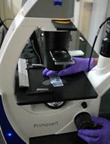
- IncuCyte ZOOM live-cell analysis system (Essen Bioscience, model: IncuCyte® ZOOM )
- FACSCanto II Flow Cytometer (BD, BD Bioscience, model: FACSCanto II )
Software
- IncuCyte ZOOM software (Essen Bioscience)
- Flow cytometry Software (Perttu Terho)
Procedure
文章信息
版权信息
© 2018 The Authors; exclusive licensee Bio-protocol LLC.
如何引用
Szalai, P. and Engedal, N. (2018). An Image-based Assay for High-throughput Analysis of Cell Proliferation and Cell Death of Adherent Cells. Bio-protocol 8(9): e2835. DOI: 10.21769/BioProtoc.2835.
分类
癌症生物学 > 细胞死亡 > 细胞生物学试验 > 细胞活性
细胞生物学 > 细胞成像 > 活细胞成像
细胞生物学 > 细胞活力 > 细胞死亡
您对这篇实验方法有问题吗?
在此处发布您的问题,我们将邀请本文作者来回答。同时,我们会将您的问题发布到Bio-protocol Exchange,以便寻求社区成员的帮助。