- EN - English
- CN - 中文
Mammalian Cell-derived Vesicles for the Isolation of Organelle Specific Transmembrane Proteins to Conduct Single Molecule Studies
哺乳动物细胞源性囊泡用于分离细胞器特异性跨膜蛋白以进行单分子研究
发布: 2018年05月05日第8卷第9期 DOI: 10.21769/BioProtoc.2825 浏览次数: 7483
评审: Rahul SrinivasanTalita Diniz Melo HanchukAnonymous reviewer(s)
Abstract
Cell-derived vesicles facilitate the isolation of transmembrane proteins in their physiological membrane maintaining their structural and functional integrity. These vesicles can be generated from different cellular organelles producing, housing, or transporting the proteins. Combined with single-molecule imaging, isolated organelle specific vesicles can be employed to study the trafficking and assembly of the embedded proteins. Here we present a method for organelle specific single molecule imaging via isolation of ER and plasma membrane vesicles from HEK293T cells by employing OptiPrep gradients and nitrogen cavitation. The isolation was validated through Western blotting, and the isolated vesicles were used to perform single molecule studies of oligomeric receptor assembly.
Keywords: Single molecule (单分子)Background
A large number of transmembrane proteins are formed through the assembly of multiple subunits leading to complicated oligomeric structures that can often exist in multiple stoichiometries. Understanding how changes in assembly alter trafficking and localization within different organelles is essential to determining a protein’s physiological role and the connection to diseases associated with maturation and transport. Single molecule approaches can provide a better understanding of the assembly of oligomeric proteins by directly measuring their stoichiometry (Ulbrich and Isacoff, 2007; Richards et al., 2012). This approach avoids ensemble averaging which provides the average state of all the stoichiometries (Walter and Bustamante, 2014). Single molecule studies have recently been employed to understand the structural and functional properties of macromolecules including conformational dynamics (Tan et al., 2014), ion channel gating (Wang et al., 2016), ligand-receptor interaction (Moonschi et al., 2015), and stoichiometric assembly (Ulbrich and Isacoff, 2007; Moonschi et al., 2015). To conduct single molecule studies, it is imperative to isolate single receptors in a supporting bilayer. Separation from the cellular environment is often necessary because the endogenous concentration of membrane proteins in mammalian cells is typically much higher than conditions compatible with single molecule studies (Richards et al., 2012). Additionally, live cell single molecule studies suffer from a number of disadvantages including high levels of auto-fluorescence, limited fluorophore brightness and the mobility of membrane proteins on the cell surface (Andersson et al., 1998; Lippincott-Schwartz et al., 1999). Isolation strategies using artificial bilayers such as liposomes are also problematic as they include an intermediate step where the protein is stabilized into a detergent solution which poses a threat to the structural integrity of the transmembrane protein. Membrane-derived vesicles enable the protein to remain embedded in its physiological membrane maintaining its structural and functional integrity. These nanoscale vesicles have been employed to study stoichiometric assembly of multimeric proteins, to probe ligand receptor interactions, and to understand the effect of ligands on the assembly of nicotinic receptors (Fox et al., 2015; Moonschi et al., 2015; Fox-Loe et al., 2017).
Transmembrane proteins are synthesized and assembled in the endoplasmic reticulum and then trafficked to the plasma membrane. Oligomeric proteins with multiple non-identical subunits can often be assembled with different subunit stoichiometries potentially leading to different trafficking and functional properties (Grady et al., 2010). For instance, α4β2 nicotinic receptors are pentameric receptor with two possible stoichiometric assemblies: (α4)2(β2)3 and (α4)3(β2)2. It has been hypothesized that cellular machinery preferentially traffics the high sensitivity isoform (α4)2(β2)3 over the other isoform from the ER to the plasma membrane. It has also been hypothesized that nicotine alters the assembly of this receptor from the low sensitivity to high sensitivity isoform in the ER leading to higher levels of the preferentially trafficked assembly in both the ER and plasma membrane (Lester et al., 2009; Henderson and Lester, 2015). Single molecule studies of proteins from specific organelles enable the correlation of changes in structural assembly of the nicotinic receptors to changes in trafficking and assembly.
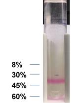
Here we present a method which can isolate single transmembrane proteins into the ER and plasma membrane derived vesicles using nitrogen cavitation and an OptiPrep gradient. The ER and plasma membrane derived vesicles exhibit different densities because they contain different phospholipids and associated proteins. ER originated vesicles are much denser than those obtained from the plasma membrane. The OptiPrep gradient was selected because of its superior ability to maintain isosmotic pressure independent of the density of the gradient used to isolate cellular organelles and subcellular vesicles (Graham et al., 1994). We applied this method to study ligand induced changes in the assembly of nicotinic receptors in the ER as well as the plasma membrane and validated our method with Western blotting and single molecule step-wise photobleaching. We believe this method can be applied for virtually any type of transmembrane proteins to conduct single molecule studies and to understand organelle-specific structural and functional properties.
Materials and Reagents
- Cell culture flasks (Greiner Bio One International, catalog number: 658175 )
- 50 ml centrifuge tubes (VWR, catalog number: 89039-658 )
- Ultra-Clear ultracentrifuge tubes (Beckman Coulter, catalog number: 344061 )
- Four 5-ml Serological pipettes (VWR, catalog number: 89130-896 )
- One 9-inch-flint-glass Pasteur pipette (VWR, catalog number: 14672-380 )
- 15 ml centrifuge tubes (VWR, catalog number: 89039-666 )
- 1.5-ml tubes
- Wine cork
- Razor blades (VWR, catalog number: 55411-050 )
- Two ice buckets (VWR, catalog number: 10146-202 )
- HEK293T cells (Sigma-Aldrich, catalog number: 85120602-1VL )
- Deionized water
- PBS buffer (VWR, catalog number: 97062-948 )
- Versene solution (Thermo Fisher Scientific, GibcoTM, catalog number: 15040066 )
- Nitrogen gas
- OptiPrep (60% solution, Sigma-Aldrich, catalog number: D1556-250ML )
- Anti-calnexin (Abcam, catalog number: ab92573 )
- Anti-sodium potassium ATPase (Abcam, catalog number: ab76020 )
- Mouse anti-Rabbit secondary antibody (Santa Cruz Biotechnology, catalog number: sc-2357 )
- ClarityTM Western ECL Substrate (Bio-Rad Laboratories, catalog number: 1705060 )
- Tris-HCl (Sigma-Aldrich, Roche Diagnostics, catalog number: 10812846001 )
- Sodium chloride (NaCl) (Fisher Scientific , catalog number: BP358-1 )
- Magnesium chloride hexahydrate (MgCl2·6H2O) (Fisher Scientific, catalog number: BP214-500 )
- Calcium chloride (CaCl2) (Fisher Scientific, catalog number: C614-500 )
- Protease inhibitor mini tablet (Thermo Fisher Scientific, Thermo ScientificTM, catalog number: A32955 )
- Sucrose (VWR, catalog number: 97063-788 )
- HEPES (Fisher Scientific, catalog number: BP310-500 )
- Hypotonic protease inhibitor solution (see Recipes)
- Sucrose buffer (see Recipes)
Equipment
- One 1 ml pipettor (Gilson, catalog number: F123602 )
- One pipet controller (VWR, catalog number: 613-4442 )
- One pair of scissors
- A set of stand (a stand and a clamp)
- Incubator
- Biosafety cabinet (Class II, Type A2)
- Centrifuge (Beckman Coulter, model: Allergra® X-22R ; Rotor: Beckman Coulter, model: SX4250 )
- Ultracentrifuge (Beckman Coulter, model: L-60 )
- Swing bucket rotor (Beckman Coulter, model: SW 28 )
- Fixed angle rotor (Beckman Coulter, model: 70 Ti )
- Belly dancer or orbital shaker
- Nitrogen cavitation chamber/vessel(Parr Instrument Company, catalog number: 4639 )
- Variable-Flow Peristaltic Pumps (Fisherbrand, Thermo Fisher)
Procedure
文章信息
版权信息
© 2018 The Authors; exclusive licensee Bio-protocol LLC.
如何引用
Moonschi, F. H., Fox-Loe, A. M., Fu, X. and Richards, C. I. (2018). Mammalian Cell-derived Vesicles for the Isolation of Organelle Specific Transmembrane Proteins to Conduct Single Molecule Studies. Bio-protocol 8(9): e2825. DOI: 10.21769/BioProtoc.2825.
分类
分子生物学 > 蛋白质 > 分离
您对这篇实验方法有问题吗?
在此处发布您的问题,我们将邀请本文作者来回答。同时,我们会将您的问题发布到Bio-protocol Exchange,以便寻求社区成员的帮助。
Share
Bluesky
X
Copy link


