

A Method for Extracting the Nuclear Scaffold from the Chromatin Network
一种从染色质网络中提取核骨架的方法
发布: 2018年04月20日第8卷第8期 DOI: 10.21769/BioProtoc.2821 浏览次数: 7600
评审: Manjula MummadisettiPearl CampbellAmey Redkar
参见作者原研究论文
The authors used this protocol in:

Feb 2018
Advertisement


Abstract
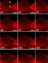
Each cell contains many large DNA polymers packed in a nucleus of approx. 10 μm in diameter. With histones, these DNA polymers are known to form chromatins. How chromatins further compact in the nucleus is unclear but it inevitably depends on an extensive non-chromatin nuclear scaffold. Imaging of endogenous chromatin network and the complementary scaffold that support this network has not been achieved but biochemical and proteomic investigations of the scaffold can still provide important insights into this chromatin-organizing network. However, this demands highly inclusive and reproducible extraction of the nuclear scaffold. We have recently developed a simple protocol for releasing the scaffold components from chromatins. The inclusiveness of the extract was testified by the observation that, upon its extraction from the nuclei, the remaining nuclear chromatins were liberated into extended and often parallel chromatin fibers. Basically, this protocol includes the generation of pure nuclei, treatment of the nuclei with Triton X-100 to generate envelope-depleted nuclei (TxN), and extraction of the nuclei at 500 mM NaCl in a sucrose-containing buffer. This combined extract of TxN is known as TxNE.
Keywords: Nuclei (核)Background
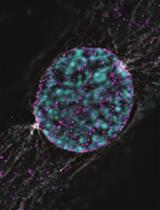
Chromatins are densely and dynamically compacted in the nuclei through a complex scaffold of proteins and ribonucleoproteins. Unlike the cytoskeletal networks (Fischer and Fowler, 2015), microscopic observation of this nuclear scaffold is technically challenging. This may reflect the dominance of chromatins inside each nucleus with which the scaffold mingles and negotiates in the nucleus. The spherical arrangement of the nucleus also contributes to challenges in imaging such scaffold structures. A major element of the nuclear scaffold is the nuclear lamina (NL) (Gruenbaum and Foisner, 2015). NL covers the surface of the entire nuclear chromatin mass on which the nuclear envelop (NE) attaches. The peripheral surface of the nuclear chromatin mass is characterized by dense heterochromatin (Gruenbaum and Foisner, 2015). Inside the nucleus, one or more nucleoli can be found that occupy significant nuclear regions (Jordan and McGovern, 1981; Shaw and Jordan, 1995; Pederson, 2011). The nucleolar surface is also surrounded by a rim of dense chromatins and potentially functions as an important interior scaffold to support the chromatins (Chen et al., 2018). The morphology of the nucleoli changes dynamically depending on the stage of the cell cycle which can significantly affect the organization of nuclear chromatins (Hernandez-Verdun, 2011; Chen et al., 2018).
The presence of such scaffold structures has been extensively investigated in 1980-90s although a direct visualization has not been achieved (Gasser, 2002; Laemmli et al., 1992). However, the composition of the nuclear scaffold and interactions among the scaffold elements could be learned through biochemical, proteomic and imaging studies. To this end, effective and reproducible extraction of the nuclear scaffold from the chromatin network is essential. We have recently developed a simple protocol for the extraction of nuclear scaffold proteins (Chen et al., 2018).
This protocol was initially developed based on a study showing that isolated nucleoli could be solubilized at 400 mM NaCl (Trimbur and Walsh, 1993). We adopted this condition to solubilize isolated nuclei. The inclusiveness of the extract was testified by the liberation of nuclear chromatins as extended parallel chromatin fibers (Chen et al., 2018). TxNE can be used to study many aspects of the nuclear scaffold.
Materials and Reagents
- 0.5-10 L pipette tips (Corning, Axygen®, catalog number: T-300 )
- 10-200 ml pipette tips (Greiner Bio One International, catalog number: 739290 )
- 100-1,000 ml pipette tip (Greiner Bio One International, catalog number: 686290 )
- T 175 cell culture flask (Thermo Fisher Scientific, Thermo ScientificTM, catalog number: 159910 )
- T 75 cell culture flasks (Thermo Fisher Scientific, Thermo ScientificTM, catalog number: 156499 )
- 150 mm circular tissue culture dishes (Thermo Fisher Scientific, Thermo ScientificTM, catalog number: 168381 )
- 50 ml Falcon tubes (Greiner Bio One International, catalog number: 227261 )
- 5 ml blunt-ended syringes (5 ml/cc, Luer slip, Cellotron, https://dcellotron.en.ec21.com/)
- 15 ml Falcon tubes (Greiner Bio One International, catalog number: 188271 )
- Nalgene Oak Ridge High-Speed Centrifuge Tubes (Thermo Fisher Scientific, Thermo ScientificTM, catalog number: 3114-0030 )
- Regular 1.5 ml microcentrifuge tubes (Corning, Axygen®, catalog number: MCT-150-C )
- Low adhesion microcentrifuge tubes (Eppendorf, catalog number: 022431081 )
- 12 mm glass coverslips (Electron Microscopy Sciences, catalog number: 72231-10 )
- Glass slides (Thermo Fisher Scientific, Thermo ScientificTM, catalog number: 6776215 )
- 50 ml syringes (Terumo, catalog number: SS*50LE )
- 0.22 μm filters (Merck, catalog number: SLGP033RS )
- HeLa S3 human cervical carcinoma cells (ATCC, catalog number: CCL-2 )
- Trypsin solution (0.05% w/v, Capricorn scientific, catalog number: TRY-1B10 )
- 0.5% Trypsin (Trypsin-EDTA, 10x stock, Thermo Fisher Scientific, GibcoTM, catalog number: 15400054 )
- Protease inhibitors PMSF (BDH, catalog number: 442172C )
- Triton X-100 (Sigma-Aldrich, catalog number: T8787 )
- 5 M NaCl
- 1% (w/v) paraformaldehyde
- Mouse anti-nucleophosmin-1 (NPM1) antibody (Sigma-Aldrich, catalog number: B0556 )
- Rabbit anti-lamin B1 (LB1) antibody (Abcam, catalog number: ab16048 )
- Rabbit anti-histone H1.2 (Abcam, catalog number: ab17677 )
- Rabbit anti-lamin A/C (Santa Cruz Biotechnology, catalog number: sc-7292 )
- Rabbit anti-H1.x antibody (Abcam, catalog number: ab31972 )
- Goat anti-mouse IgG (Cy3-conjugated) (Jackson ImmunoResearch Laboratories, catalog number: 115-165-003 )
- Goat anti-rabbit IgG (Alexa Fluor 488-conjugated) (Jackson ImmunoResearch Laboratories, catalog number: 115-545-003 )
- Mounting medium (Vector laboratories, catalog number: H-1200 )
- Any clear Nail polish (We use Daiso Japan Nail Top Coat Clear, 10 ml bottle, made in Taiwan)
- 12.5% (w/v) gels
- Sodium chloride (NaCl) (Merck)
- Potassium chloride GR (KCl)(Merck)
- Di-sodium hydrogen phosphate (Merck)
- Sucrose (First BASE Laboratories, catalog number: BIO-1090-1kg )
- Sucrose (Merck)
- Tris (Merck)
- Tris base (Avantor Performance Materials, J.T. Baker, catalog number: 4109-02 )
- Magnesium chloride hexahydrate (Merck, catalog number: 1058331000 )
- Magnesium chloride hexahydrate (MgCl2·H2O)( Merck)
- DMEM medium (Thermo Fisher Scientific, GibcoTM, catalog number: 12430054 )
- Fetal bovine serum (FBS, GE Healthcare, HyClone, catalog number: SV30160.03 )
Note: We purchased FBS and generated heat-inactivated FBS by incubation at 56 °C for 30 min. - Penicillin-Streptomycin (100x stock, PAN Biotech, catalog number: P06-07100 )
- L-glutamine (200 mM, 100x stock, Thermo Fisher Scientific, GibcoTM, catalog number: 25030081 )
- PBS, pH 7.4 (see Recipes)
- 0.25 M sucrose buffer, pH 7.4 (see Recipes)
- 2.2 M sucrose buffer, pH 7.4 (see Recipes)
- DMEM culture medium (see Recipes)
Equipment
- 1,000 L pipette (Eppendorf, model: Research® plus, catalog number: 3120000062 )
- 200 L pipette (Eppendorf, model: Research® plus, catalog number: 3120000054 )
- Humidified cell incubator (Thermo Fisher Scientific, Thermo ScientificTM, model: FormaTM Steri-CycleTM CO2 incubator , catalog number: 371)
- Homogenizer (Isobiotec Precision Engineering, Heidelberg, Germany)
- Tabletop microcentrifuge (Beckman Coulter, model: Microfuge 22R centrifuge , catalog number: 368831)
- Tabletop swing bucket centrifuge (Eppendorf, model: 5810 R , catalog number: 5811000320)
- Beckman Coulter centrifuge with a JA-20 rotor (Beckman Coulter, model: Avanti J-25 , catalog number: 363102)
- BSL-2 cell culture cabinet (Gelman, model: Bioessential Class II series Type A2 Laminar flow BSC, http://gelmansingapore.com/product/bioessential-class-ii-type-a2-series-laminar-flow-biological-safety-cabinets/)
- FluoView FV1000 confocal microscope
- Cool/SNAP HQ2 image acquisition camera (Olympus)
Software
- FV-ASW 1.6b software
- Imaris software (Bitplane AG)
Procedure
文章信息
版权信息
© 2018 The Authors; exclusive licensee Bio-protocol LLC.
如何引用
Readers should cite both the Bio-protocol article and the original research article where this protocol was used:
- Chen, J., Teo, B. H. D. and Lu, J. (2018). A Method for Extracting the Nuclear Scaffold from the Chromatin Network. Bio-protocol 8(8): e2821. DOI: 10.21769/BioProtoc.2821.
- Chen, J., Teo, B. H. D., Cai, Y., Wee, S. Y. K. and Lu, J. (2018). The linker histone H1.2 is a novel component of the nucleolar organizer regions. J Biol Chem 293(7): 2358-2369.
分类
细胞生物学 > 细胞结构 > 细胞核
您对这篇实验方法有问题吗?
在此处发布您的问题,我们将邀请本文作者来回答。同时,我们会将您的问题发布到Bio-protocol Exchange,以便寻求社区成员的帮助。