

Ciliary Assembly/Disassembly Assay in Non-transformed Cell Lines
非转化细胞系中睫毛的组装/解聚测定
(*contributed equally to this work) 发布: 2018年03月20日第8卷第6期 DOI: 10.21769/BioProtoc.2773 浏览次数: 10366
评审: Ralph Thomas BoettcherChris TibbittMurugappan Sathappa
参见作者原研究论文
The authors used this protocol in:

Aug 2017


Abstract
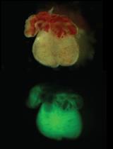
The primary cilium is a non-motile sensory organelle whose assembly and disassembly are closely associated with cell cycle progression. The primary cilium is elongated from the basal body in quiescent cells and is resorbed as the cells re-enter the cell cycle. Dysregulation of ciliary dynamics has been linked with ciliopathies and other human diseases. The in vitro serum-stimulated ciliary assembly/disassembly assay has gained popularity in addressing the functions of the protein-of-interest in ciliary dynamics. Here, we describe a well-tested protocol for transfecting human retinal pigment epithelial cells (RPE-1) and performing ciliary assembly/disassembly assays on the transfected cells.
Keywords: Primary cilium (初级纤毛)Background
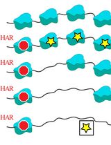
Primary cilia are hair-like sensory organelles that appear at the G0/G1 phase, and are disassembled prior to the S phase of the cell cycle (Tucker et al., 1979). Previous studies have confirmed that certain non-transformed cell types (i.e., RPE-1 cells, 3T3 fibroblasts, and mouse embryonic fibroblasts [MEFs]) can be starved to induce quiescence and ciliary formation. Subsequent re-addition of serum triggers biphasic ciliary resorption, which peaks at 2 h and 24 h following stimulation (Tucker et al., 1979; Li et al., 2011). This phenomenon lays the foundation for the serum-stimulated ciliary assembly/disassembly assay commonly used in the literature to identify proteins involved in ciliary assembly and disassembly (Pugacheva et al., 2007; Saito et al., 2017). MEFs derived from transgenic mice (in which the gene-of-interest is deleted) are often used to investigate the dynamic role of a given protein in the ciliary assembly/disassembly assays. When the specific MEF types are not accessible, one may modify the expression level of the targeted protein in naive RPE-1 cells (or 3T3 or MEFs) using transfection of cDNA or short hairpin RNA. We describe the protocol of these procedures that are routinely carried out in the lab. To unambiguously identify the cell autonomous effect on ciliary assembly or disassembly in the transfected cells, we typically ‘tag’ the transfected cells with green fluorescence via the expressed GFP or GFP-fusion protein.
Materials and Reagents
- 10 µl pipette tips (Corning, catalog number: 4110 , or Denville Scientific, catalog number: P1096 )
- 200 µl pipette tips (FUKAEKASEI and WATSON, catalog number: 110-705Y , or Denville Scientific, catalog number: P1122 )
- 1,000 µl pipette tips (FUKAEKASEI and WATSON, catalog number: 110-706B , or Denville Scientific, catalog number: P2103-N )
- 100 mm cell culture dish (AS ONE, Violamo, catalog number: 2-8590-03 , or Corning, catalog number: 430167 )
- 1.5 ml microcentrifuge tube (Sorenson BioScience, catalog number: 11510 , or National Scientific, catalog number: CN1700-BP )
- Precleaned, sterile 12-13 mm micro-glass coverslips (Matsunami Glass, catalog number: C013001 , or Thermo Fisher Scientific, catalog number: 12CIR-1.5 )
- 35 mm cell culture dish (Corning, Falcon®, catalog number: 353001 , or Corning, catalog number: 430165 )
- 15 ml centrifuge tube (FUKAEKASEI and WATSON, catalog number: 1332-015S , or Corning, catalog number: 430053 )
- 24-well plate (NuncTM, Thermo Fisher Scientific, Thermo ScientificTM, catalog number: 142475 , or Corning, catalog number: 3527 )
- Parafilm
- 14 x 16 cm glass (or plastic) plates
- Micro-slide glass (Matsunami Glass, catalog number: S024410 )
- 0.22 µm filter (Corning, catalog number: 431097 , or Advantec MFS, catalog number: 25CS020AS )
- RPE-1 cells (ATCC, catalog number: CRL-4000 )
- Midi-prepared, or maxi-prepared plasmid (> 1 µg/µl is preferred)
Note: We typically have the cDNA (under the CAG or CMV promoter) or short hairpin RNA (shRNA; under the U6 promoter) inserted in the same plasmid that also expresses GFP (e.g., pCAGIG vector). - 0.05% Trypsin-0.53 mM EDTA (Wako Pure Chemical Industries, catalog number: 202-16931 )
- Fetal bovine serum (FBS) (PAA Laboratories, catalog number: A15-701 )
- Dulbecco’s modified Eagle medium/Ham’s F-12 (DMEM/F12) (Wako Pure Chemical Industries, catalog number: 048-29785 )
- Methanol
- Monoclonal anti-acetylated α-tubulin (Ac-Tub) antibody, clone 6-11B-1 (Sigma-Aldrich, catalog number: T6793 )
- Anti-Tubulin Antibody, Detyrosinated (Merck, catalog number: AB3201 )
- Monoclonal anti-γ-tubulin antibody, clone GTU-88 (Sigma-Aldrich, catalog number: T5326 )
- Polyclonal anti-GFP tag antibody (Thermo Fisher Scientific, InvitrogenTM, catalog number: A-6455 )
- Goat anti-mouse IgG (H+L) antibody, Alexa Fluor 568 (Thermo Fisher Scientific, InvitrogenTM, catalog number: A-11004 )
- Goat anti-mouse IgG2b antibody, Alexa Fluor 568 (Thermo Fisher Scientific, InvitrogenTM, catalog number: A-21144 )
- Goat anti-mouse IgG1 antibody, Alexa Fluor 647 (Thermo Fisher Scientific, InvitrogenTM, catalog number: A-21240 )
- Goat anti-Rabbit IgG (H+L) antibody, Alexa Fluor 488 (Thermo Fisher Scientific, InvitrogenTM, catalog number: R37116 )
- Fluorescent mounting medium (Agilent Technologies, Dako, catalog number: S302380-2 , code number: S3023) or ProLong Gold Antifade Mountant (Thermo Fisher Scientific, InvitrogenTM, catalog number: P36930 )
- Nail polish
- Dulbecco’s modified Eagle medium (DMEM) (NACALAI TESQUE, catalog number: 08458-16 )
- 100 mM pyruvate (Thermo Fisher Scientific, GibcoTM, catalog number: 11360070 )
- 10x D-PBS (-) (Wako Pure Chemical Industries, catalog number: 048-29805 )
- Calcium chloride dihydrate (CaCl2·2H2O) (Wako Pure Chemical Industries, catalog number: 031-00435 )
- Magnesium chloride hexahydrate (MgCl2·6H2O) (Wako Pure Chemical Industries, catalog number: 135-00165 )
- 16% Paraformaldehyde (PFA) aqueous solution (Electron Microscopy Science, catalog number: 15710 )
- Ammonium chloride (NH4Cl) (Wako Pure Chemical Industries, catalog number: 017-02995 )
- Bovine serum albumin (BSA) (Wako Pure Chemical Industries, catalog number: 010-15114 )
- Triton X-100 (GE Healthcare, catalog number: 17-1315-01 )
- Sodium azide (NaN3) (Wako Pure Chemical Industries, catalog number: 190-01272 )
- 4’,6-Diamidino-2-phenylindole (DAPI) (Thermo Fisher Scientific, InvitrogenTM, catalog number: D1306 )
- Growth medium (see Recipes)
- PBS for cell culture (see Recipes)
- 2x PBSc/m (see Recipes)
- PBSc/m (see Recipes)
- 4% PFA/PBSc/m (see Recipes)
- 50 mM NH4Cl (see Recipes)
- BTPAD (see Recipes)
Equipment
- 5% CO2/95% air tissue culture incubator (SANYO, model: MCO-18AIC )
- Water bath (TOKYO RIKAKIKAI, Eyela, model: NTT-1200 )
- Hemocytometer chamber (Erma, catalog number: 03-303-1 )
- Low-speed centrifuge (TOMY SEIKO, model: LC-100 ; Eppendorf, model: 5702 )
- Pipette 20 µl (Pipetman P) (Gilson, catalog number: F123600 )
- Pipettes 200 µl (Pipetman P) (Gilson, catalog number: F123601 )
- Pipettes 1,000 µl (Pipetman P) (Gilson, catalog number: F123602 )
- For Procedure B1:
NeonTM transfection system (Thermo Fisher Scientific, InvitrogenTM, catalog number: MPK5000 ) - For Procedure B2:
- NucleofectorTM device (Lonza, model: NucleofectorTM I )
- Cuvettes PlusTM Electroporation Cuvettes & transfer tips (BTX, catalog number: 45-0135 )
- Amaxa® Cell Line Nucleofector® Kit V (Lonza, catalog number: VCA-1003 )
- NucleofectorTM device (Lonza, model: NucleofectorTM I )
- Forceps (Fine Scientific Tool, Dumont, model: #5/45 )
- Autoclave (TOMY SEIKO, model: LBS-245 )
- Epifluorescent microscope (Carl Zeiss, model: Axioplan 2 imaging ) equipped with objective lens (EC Plan-Neofluar 40x/0.75, Carl Zeiss, catalog number: 440350-9903-000 )
- Confocal microscope (ZEISS, model: LSM-780 )
Procedure
文章信息
版权信息
© 2018 The Authors; exclusive licensee Bio-protocol LLC.
如何引用
Saito, M., Sakaji, K., Otsu, W. and Sung, C. (2018). Ciliary Assembly/Disassembly Assay in Non-transformed Cell Lines. Bio-protocol 8(6): e2773. DOI: 10.21769/BioProtoc.2773.
分类
发育生物学 > 细胞生长和命运决定 > 增殖
发育生物学 > 细胞信号传导 > 配体
细胞生物学 > 基于细胞的分析方法 > 细胞器能动性
您对这篇实验方法有问题吗?
在此处发布您的问题,我们将邀请本文作者来回答。同时,我们会将您的问题发布到Bio-protocol Exchange,以便寻求社区成员的帮助。