

Organotypic Explants of the Embryonic Rodent Hippocampus: An Accessible System for Transgenesis
啮齿动物胚胎海马器官型外植体:一个可进行转基因的系统

(*contributed equally to this work) 发布: 2018年03月20日第8卷第6期 DOI: 10.21769/BioProtoc.2764 浏览次数: 7640
评审: Oneil G. BhalalaAnonymous reviewer(s)
参见作者原研究论文
The authors used this protocol in:

Jul 2011
Advertisement


Abstract
This protocol describes the technique of ex-vivo electroporation to target embryonic hippocampal progenitors in an organotypic slice preparation. This technique allows gene perturbation for examining developmental processes in the embryonic hippocampus while retaining the environment and connectivity of the cells. Gene perturbation can include Cre-mediated recombination, RNAi-mediated knockdown, gene overexpression, or a combination of any of these. Ex-vivo electroporation can be performed at a wide range of embryonic stages, giving temporal control to the experimenter. Spatial control can be achieved more easily by preparing the brain in a Petri dish to target particular regions of the hippocampus. The electroporated explant cultures provide a highly tractable system for the study of developmental processes that include progenitor proliferation, migration and cell fate acquisition.
Keywords: Mouse hippocampus (小鼠海马)Background
The hippocampus presents a challenge in terms of accessibility due to its location in the caudomedial telencephalon. The embryonic hippocampus is even more inaccessible, requiring in utero surgical methods for experimental manipulation. Organotypic slice cultures circumvent this problem and at the same time retain many aspects of hippocampal field cytoarchitectonics, including molecular features and connectivity. While there are protocols that describe postnatal culturing of hippocampal explants from rodent brains (Stoppini et al., 1991; Opitz-Araya and Barria, 2011) these do not include genetic manipulation of the cells. The preparation of organotypic explants from the embryonic mouse hippocampus was first described in Tole et al. (1997). We extended this protocol by introducing ex-vivo electroporation of the embryonic brain prior to preparing the organotypic slices. Electroporation of the intact brain after introducing DNA into the telencephalic ventricle ensures that cells residing in and near the ventricular zone are targeted, and therefore provides an excellent means of inducing transgenesis in hippocampal progenitors. Data using this protocol were published in Subramanian et al. (2011). Here, we present detailed step-wise instructions including experimental ‘dos and don’ts’, and also illustrate key steps using photographs and movies, to aid new researchers in setting up this useful procedure.
Some advantages and applications of this protocol are:
1)Temporal control can be achieved by isolating the embryonic hippocampus at the desired stage to access early, mid, or late-gestation progenitors.
2)Spatial control can be achieved by orienting the electrodes to target the hippocampus or different areas of the cortex.
3)Cre-mediated recombination can be employed by electroporating Cre-GFP into embryos carrying the desired floxed alleles.
4)Overexpression constructs can be electroporated.
5)Embryonic lethal strains can be accessed by performing the procedure in the window of viability, and then further development can proceed in the organotypic explant.
Materials and Reagents
- Plastic Pasteur pipettes:
3 ml (P-3) and 2 ml (P-1) (Taurus Biomedical)
1.5 ml pipettes (RPI, catalog number: 147500 )
Note: Henceforth these will all be referred to as ‘pipettes’. Procuring the correct size of pipette and cutting the shaft to the correct aperture size (see Figure 1) is key to being able to manipulate brains, hemispheres and explants without damaging them.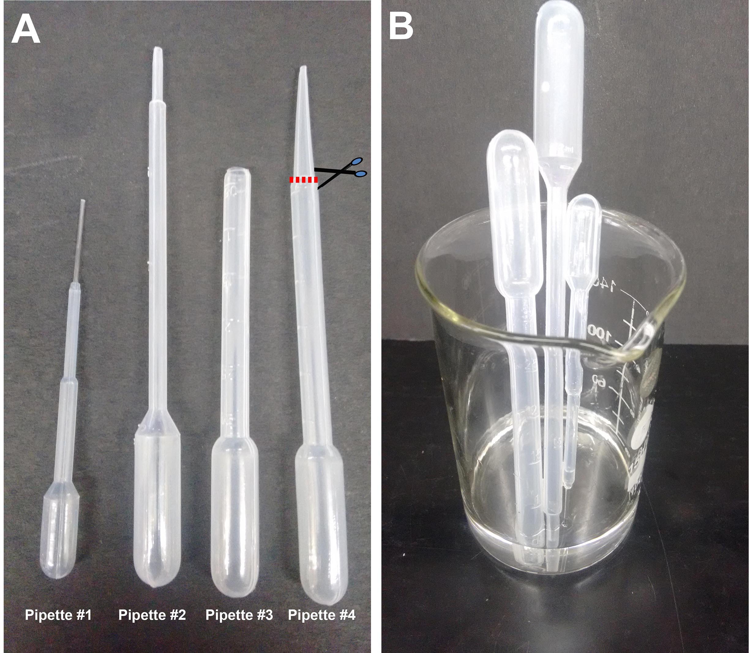
Figure 1. Plastic Pasteur pipettes. A. Different sizes of pipettes used in this procedure: #1 (1.5 ml pipette), #2 (2 ml pipette), #3 (3 ml pipette) prepared by cutting #4 as shown. The cut is made just at the point where the pipette shaft diameter begins to taper, approximately 2 cm from the tip. B. Beaker with pipettes being sterilized in alcohol. - 35 mm sterile Petri dishes (Laxbro, catalog number: PD-35 TC )
- Cell culture 6-well tissue culture plate (Thermo Fisher Scientific, catalog number: 140675 )
- Micropipette tip
- Glass Capillaries: Thin wall borosilicate tubing without filament (Sutter Instruments, catalog number: B100-75-10 )
- 100 mm Petri dishes (Thermo Fisher Scientific, Thermo ScientificTM, catalog number: 150464 )
- Sterile blade
- Cell Culture Inserts: 0.4 µm (Millicell, Merck, catalog number: PICM03050 )
- Aspirator assembly (Sigma-Aldrich, catalog number: A5177-5EA )
- 50 ml syringe filter unit with 0.22 µm filter (Millex-GP, Merck, catalog number: SLGP033RS ) for sterilizing the culture medium
- 2 Squirt bottles (Tarsons, catalog number: 561100 ) (containing 70% and 100% ethanol)
- pCS2-EGFP plasmid (or any desired plasmid)
- Pregnant Swiss mice (SWR/J) (obtained from Tata Institute of Fundamental Research breeding facility)
- Absolute ethanol (Merck, catalog number: 107017 )
- Leibovitz’s L-15 medium (Thermo Fisher Scientific, GibcoTM, catalog number: 41300039 )
- Fast green dye (Sigma-Aldrich, catalog number: F7252 )
- Penicillin-streptomycin (P/S) (10,000 U/ml) (Thermo Fisher Scientific, GibcoTM, catalog number: 15140122 )
- Dulbecco’s modified Eagle medium (Thermo Fisher Scientific, GibcoTM,catalog number: 12800017 )
- B27TM supplement (50x), serum-free (Thermo Fisher Scientific, GibcoTM, catalog number: 17504044 )
- Working solution of DMEM medium (see Recipes)
Equipment
- Horizontal flow hood (Kirloskar, Envair Electrodyne, catalog number: KCH-B )
- Glass beaker (Borosil, catalog number: 1000D21 )
- Tissue chopper (McIlwain, model: MTC/2E )
- Stereo microscope (Olympus, catalog number: SZ61 ) placed in a horizontal flow tissue culture hood
- Cell culture incubator (Thermo Fisher Scientific, model: HeracellTM 150 )
- Fine tools for manipulating embryonic brains:
- Coarse forceps (Roboz Surgical Instrument, catalog number: RS-5040 ) for handling the chopper stage disc
- Fine scissors (Fine Science Tools, catalog number: 14060-11 ) and tissue forceps (Roboz Surgical Instrument, catalog number: RS-8166 ) for opening the dam and removing the uterus
- Electrodes (3 mm, BTX, Harvard Apparatus, catalog number: 450487 )
- Electroporator (BTX, Harvard Apparatus, model: ECM 830 )
- Dual-stage glass micropipette puller (NARISHIGE, model: PC-10 )
Procedure
文章信息
版权信息
© 2018 The Authors; exclusive licensee Bio-protocol LLC.
如何引用
Iyer, A., Subramanian, L. and Tole, S. (2018). Organotypic Explants of the Embryonic Rodent Hippocampus: An Accessible System for Transgenesis. Bio-protocol 8(6): e2764. DOI: 10.21769/BioProtoc.2764.
分类
神经科学 > 发育 > 外植体培养
细胞生物学 > 组织分析 > 电穿孔
细胞生物学 > 组织分析 > 组织分离
您对这篇实验方法有问题吗?
在此处发布您的问题,我们将邀请本文作者来回答。同时,我们会将您的问题发布到Bio-protocol Exchange,以便寻求社区成员的帮助。