

Mouse Phrenic Nerve Hemidiaphragm Assay (MPN)
小鼠中膈神经偏侧膈分析(MPN)


发布: 2018年03月05日第8卷第5期 DOI: 10.21769/BioProtoc.2759 浏览次数: 10561
评审: Andrea PuharJordi MolgóAnonymous reviewer(s)
参见作者原研究论文
The authors used this protocol in:

Aug 2017
Advertisement


Abstract
The neuromuscular junction (NMJ) is the specialized synapse by which peripheral motor neurons innervate muscle fibers and control skeletal muscle contraction. The NMJ is the target of several xenobiotics, including chemicals, plant, animal and bacterial toxins, as well as of autoantibodies raised against NMJ antigens. Depending on their biochemical nature, the site they target (either the nerve or the muscle) and their mechanism of action, substances affecting NMJ produce very specific alterations of neuromuscular functionality.
Here we provide a detailed protocol to isolate the diaphragmatic muscle from mice and to set up two autonomously innervated hemidiaphragms. This preparation can be used to study bioactive substances like toxins, venoms and neuroactive molecules of various origin, or to measure the force of skeletal muscle contraction.
The ‘mouse phrenic nerve hemidiaphragm assay’ (MPN) is an established model of ex vivo NMJ and recapitulates the complexity of neuromuscular transmission in a system easy to control and to manipulate, thus representing a valuable tool to study both NMJ physiology and the mechanism of action of toxins and other molecules acting at this synapse.
Background
The neuromuscular junction (NMJ) is the chemical synapse enabling communication between motor neurons and skeletal muscle fibers. This is the best characterized synapse and most of the knowledge on maturation, structure and function of synapses derives from its study (Li et al., 2017). At the NMJ, the action potential running along the motor axon invades the nerve terminal (presynaptic bouton) and induces the fusion of synaptic vesicles with the presynaptic membrane. This triggers the release of acetylcholine (ACh), the neurotransmitter that binds the nicotinic ionotropic ACh receptors (nAChRs) on the postsynaptic muscle fiber. Upon ACh binding to nAChRs, a postsynaptic action potential spreads out along the muscle fiber causing Ca2+ release from the sarcoplasmic reticulum into the cytosol, thereby inducing muscle contraction. At variance from central synapses, NMJs are not protected by anatomical barriers, like the blood brain barrier or the blood nerve barrier, and are exposed to the action of various pathogenic molecules, including chemicals, toxins from plant, animal and bacteria as well as autoantibodies raised against NMJ antigens. Depending on the way they act, these agents produce distinct kinds of injury to the NMJ, eventually leading to impairment of muscle contraction.
The ‘mouse phrenic nerve hemidiaphragm assay’ (MPN) is an established model to study ex vivo NMJ function and offers a valuable tool to investigate the mechanism of action of toxins and molecules that produce NMJ alteration. In addition, MPN can be used to evaluate skeletal muscle contractility and measure diaphragmatic muscle force elicited by electrical stimulation of its phrenic nerve, so providing a model which recapitulates the complexity of the neuromuscular system in a more accessible and isolated environment.
The MPN provided fundamental insights to tackle the mechanism of action of Botulinum Neurotoxins (BoNTs), and it is currently used in many laboratories worldwide to perform qualitative/quantitative analysis of BoNTs. Its employment allows a significant refinement and reduction in the use of animals and results are in good agreement with the classical mouse lethality bioassay (Rasetti-Escargueil et al., 2011; Bigalke and Rummel, 2015).
For this, and because the hemidiaphragm-phrenic nerve intoxicated by BoNTs closely mimics the failure of respiratory muscles occurring in vivo, the MPN is presently listed in the European Pharmacopeia as an alternative method to the mouse bioassay for assaying BoNT/A lots for human use (https://ntp.niehs.nih.gov/iccvam/suppdocs/feddocs/eur/eph_botat-508.pdf). This system is also a valuable tool to test new BoNTs (Zanetti et al., 2017) and the neutralizing potency of antibodies and inhibitors (Rasetti-Escargueil et al., 2011; Azarnia Tehran et al., 2015; Beske et al., 2017).
Besides BoNTs, the MPN can be used to study many other bioactive substances, comprising toxins like phospholipases, myotoxins and complete venoms, neuroactive molecules like peptides, lipids and drugs (Rigoni et al., 2005; Caccin et al., 2006; Bercsenyi et al., 2014; Yan et al., 2014; Caccin et al., 2015), or to measure muscle force in diaphragms from mice pre-treated with autoantibodies involved in myasthenic syndromes (Klooster et al., 2012) or from animal models of neuromuscular diseases (Nascimento et al., 2014).
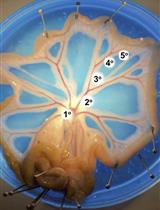
Here, we describe a very detailed protocol to successfully dissect the mouse diaphragmatic muscle with both the phrenic nerves completely functional. This is a remarkable advantage as it allows obtaining two autonomously innervated hemidiaphragms to be independently used, either as an internal control or to increment the number of experimental data. The small volume of muscle bath-chambers, the possibility of finely control bath concentration of substances used, the easy manipulability of experimental conditions (temperature, washes, etc.) and the possibility to use the muscles for further analysis (immunofluorescence, Western blot, etc.), represent significant advantages as well.
Materials and Reagents
- 2 µl micropipette
- 200 µl micropipette
- 1,000 µl micropipette
- Tips for 2, 200 and 1,000 µl micropipettes
- Petri dish, 100 x 20 mm, coated with Sylgard (Dow Corning, Sylgard® 184 Silicone Elastomer kit)
- Surgical needles (Rudolf, catalog number: RU 5899-01 )
- Cotton thread
- Mice of desired genotype and age
- Hydrogen chloride (HCl) (Sigma-Aldrich, catalog number: H1758 )
- Sodium bicarbonate (NaHCO3) (Sigma-Aldrich, catalog number: S5761 )
- Potassium chloride (KCl) (Sigma-Aldrich, catalog number: P9333 )
- Potassium phosphate monobasic (KH2PO4) (Sigma-Aldrich, catalog number: P5655 )
- Sodium chloride (NaCl) (Sigma-Aldrich, catalog number: S3014 )
- Magnesium chloride, standard solution 1 M (MgCl2) (Honeywell International, Fluka, catalog number: 63020 )
- Calcium chloride dihydrate (CaCl2·2H2O) (Sigma-Aldrich, catalog number: C5080 )
- D-(+)-Glucose (Sigma-Aldrich, catalog number: G7528 )
- Ringer’s solution (see Recipes)
Equipment
- Volumetric flask
- Clamp (Bulldog Clamp, Diethrich) (Rudolf, catalog number: RU 3934-16 ; or similar)
- 2 x scissors (Delicate Surgical Scissors) (Rudolf, catalog number: RU 1503-12 )
- 2 x forceps (Micro Jewelers Forceps, curved) (Rudolf, catalog number: RU 4240-07 )
- Stimulator: 6002 Stimulator (Harvard Apparatus, model: 6002 Basic Stimulator )
- 2 x micromanipulator (three-dimensional coarse manual manipulator) (NARISHIGE, catalog number: M-152 )
- 2 x Tension transducer: isometric transducer (Harvard Apparatus, catalog number: 72-4494 )
- 2 x Lead Screw Positioner (Harvard Apparatus, catalog number: 53-2082W )
- 2 x Support system (to hold both stimulation chamber and transducer sensors)
- 2 x Static stimulation chamber (jacketed, total internal volume 5 ml)
- Thermostatic bath (Isco, catalog number: GTR190 )
- Recording unit: i-WORX 118 system (iWORX, model: iWORX 118 )
- Computer compatible with the software
- Gas tank (95% O2, 5% CO2) equipped with pressure control (Air Liquide, model: HBS 240-1-2 )
Software
- Recording and Analysis: i-WORX 118 system (Dover, NH, USA) interfaced via Labscribe software (iWorx Systems Inc., Dover, NH, USA)
Procedure
文章信息
版权信息
© 2018 The Authors; exclusive licensee Bio-protocol LLC.
如何引用
Zanetti, G., Negro, S., Pirazzini, M. and Caccin, P. (2018). Mouse Phrenic Nerve Hemidiaphragm Assay (MPN). Bio-protocol 8(5): e2759. DOI: 10.21769/BioProtoc.2759.
分类
神经科学 > 周围神经系统 > 骨骼肌
细胞生物学 > 细胞信号传导 > 突触传递
细胞生物学 > 组织分析 > 电生理学
您对这篇实验方法有问题吗?
在此处发布您的问题,我们将邀请本文作者来回答。同时,我们会将您的问题发布到Bio-protocol Exchange,以便寻求社区成员的帮助。