

Preparation of Precisely Oriented Cryosections of Undistorted Drosophila Wing Imaginal Discs for High Resolution Confocal Imaging
制备未畸变果蝇翅成虫盘精确定向冷冻切片以用于高分辨率共焦成像

发布: 2018年02月05日第8卷第3期 DOI: 10.21769/BioProtoc.2725 浏览次数: 9913
评审: Jihyun KimImre GáspárAnonymous reviewer(s)
参见作者原研究论文
The authors used this protocol in:

Jul 2019
Advertisement


Abstract
The combination of immunofluorescence and laser scanning confocal microscopy (LSM) is essential to high-resolution detection of molecular distribution in biological specimens. A frequent limitation is the need to image deep inside a tissue or in a specific plane, which may be inaccessible due to tissue size or shape. Recreating high-resolution 3D images is not possible because the point-spread function of light reduces the resolution in the Z-axis about 3-fold, compared to XY, and light scattering obscures signal deep in the tissue. However, the XY plane of interest can be chosen if embedded samples are precisely oriented and sectioned prior to imaging (Figure 1). Here we describe the preparation of frozen tissue sections of the Drosophila wing imaginal disc, which allows us to obtain high-resolution images throughout the depth of this folded epithelium.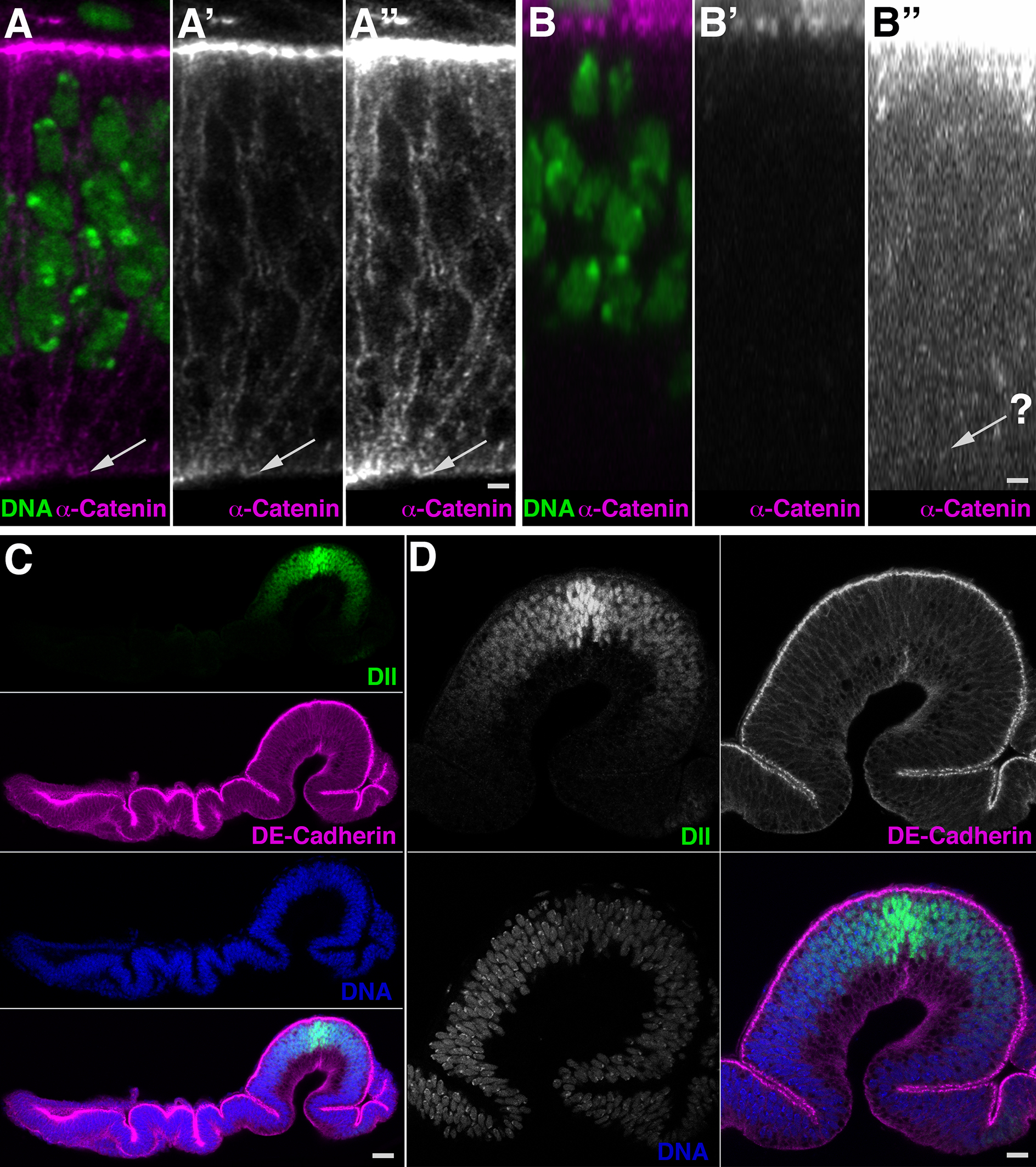
Figure 1. The epithelial structure and undistorted folding pattern are revealed in its entire depth in this frozen section of developing Drosophila wing. A-D. Transverse dorsoventral sections through the wing pouch. A. Cryosection reveals nuclei (A, green) and subcellular distribution of α-catenin (A’, A”, magenta) with signal throughout the depth of the epithelium. The basal surface is clearly detectable (arrows). A” is digitally enhanced image of A’. B. A Z-stack of images collected in a top-down view displayed as XZ orthogonal view reveals nuclei (B) but little discernable detail for α-catenin (B’, B”) and even the digitally enhanced image (B”) fails to reveal the basal epithelial surface (arrow). C. Transverse dorsoventral section displaying the Distal-less (Dll, green) gradient in the wing pouch and subcellular localization of DE-Cadherin (magenta) throughout the epithelium. D. View of the wing pouch. Dorsal is to the left; apical is up. Scale bars are 1 µm in A, B, 11 µm in C, 5 µm in D.
Background
Third instar imaginal discs are flat pocket-like involutions of the epidermis (Cohen, 1993; McClure and Schubiger, 2005). One layer of this pocket, the ‘disc proper’, is a pseudostratified columnar epithelium that is heavily folded at the onset of metamorphosis. It is continuous with the ‘overlaying’ squamous epithelium, the peripodial membrane. The focus of our work is to understand how the Wnt morphogen patterns the dome-shaped wing pouch region of the wing disc. As imaginal discs are flat overall, conventional imaging has them mounted for top-down or upside-down observation, whereby cover-slips compress and distort the folded structure. The use of spacers prevents distortions, but imaging of the entire wing pouch using Z-stacks has proved unsatisfactory or impossible; as outlined above, the reduced resolution in the Z-axis typically prevents high-resolution reconstruction of the epithelium in the apical/basal direction. Therefore, only the apical half of the epithelium of the wing pouch is detected at high resolution. This problem is exacerbated if weak signals are to be detected. Thus, uniform imaging requires a ‘side-view’ that can be obtained in sections. We modified a cryosection protocol (Culbertson et al., 2011; Sui et al., 2012) to obtain transverse sections of wing discs at defined angles. This methodology was critical to our analysis of signaling gradients in the wing pouch.
Materials and Reagents
- Pipette tips (USA Scientific, catalog numbers: 1111-1800 , 200 µl; 1111-2821 , 1,000 µl)
- Glass 9-well plate (PYREXTM Spot Plate, Fisher Scientific, catalog number: 13-748B )
- Razor blades (Single Edge Razor Blades, Stanley Black & Decker, catalog number: 28-510 )
- Conical tipped plastic embedding capsules (Electron Microscopy Sciences, BEEM®, catalog number: 69913-01 )
- Glass microscope slides (FisherfinestTM Premium Frosted, Fisher Scientific, catalog number: 12-544-2 )
- Microscope cover glass (coverslips), 22 x 50-1 (Fisher Scientific, Fisherbrand, catalog number: 12-545E )
- Plastic cling wrap
- Late third instar Drosophila melanogaster larvae
- Tissue Freezing Medium (TFMTM), clear (General Data, catalog number: TFM-C )
- Fluoromount-G® (SouthernBiotech, catalog number: 0100-01 )
- Clear Nail polish (i.e., Sally Hansen Hard As Nails)
- Sodium chloride (NaCl) (Fisher Scientific, catalog number: S271 )
- Potassium chloride (KCl) (Fisher Scientific, catalog number: P330 )
- Sodium bicarbonate (NaHCO3) (Fisher Scientific, catalog number: S233 )
- Calcium chloride dihydrate (CaCl2·2H2O) (Fisher Scientific, catalog number: C79 )
- Sodium phosphate dibasic anhydrous (Na2HPO4) (Fisher Scientific, catalog number: S374 )
- Potassium phosphate monobasic (KH2PO4) (Fisher Scientific, catalog number: P285 )
- Triton X-100 (Sigma-Aldrich, catalog number: X100 )
- Normal goat serum (Jackson ImmunoResearch, catalog number: 005-000-121 )
- Gelatin from porcine skin (gel strength 300 Type A, Sigma-Aldrich, catalog number: G2500 )
- D-Sucrose (Fisher Scientific, catalog number: BP220 )
- Sodium azide (Fisher Scientific, catalog number: S2271 )
- 16% paraformaldehyde (Ted Pella, catalog number: 18505 )
- Ice
- Dry ice
- Ringer’s solution (see Recipes)
- 4% formaldehyde fix (Solution A; see Recipes)
- 10x phosphate-buffered saline (PBS) (see Recipes)
- PBT (see Recipes)
- 5% normal goat serum (Solution B; see Recipes)
- 30% sucrose (Solution C; see Recipes)
- 10% gelatin (Solution D; see Recipes)
Equipment
- Dissection microscope (Leica Biosystems, model: Leica MZ6 )
- Fine forceps (Dumont Tweezers #5, World Precision Instruments, catalog number: 500085 )
- Pipettes 20 µl, 200 µl, 1,000 µl (Gilson, model: Pipetman P20, catalog number: F123600 ; Gilson, model: Pipetman P200, catalog number: F123601 ; Gilson, model: Pipetman P1000, catalog number: F123602 )
- Water bath, 50 °C (Fisher Scientific, model: IsotempTM 205 )
- Cryostat (Leica Biosystems, model: Leica CM1850 , with anti-roll plate assembly, Leica Biosystems, catalog number: 14041933981 )
Note: The product “Leica CM1850” has been discontinued. - Sample holders for cryostat (Specimen disc, 25 mm, Leica, catalog number: 14041619275 )
- Ultralow Temperature Freezer, -80 °C (Thermo Fisher Scientific, model: UXF60086A , catalog number: 315673H01)
- Fluorescence/confocal microscope (laser scanning microscope, ZEISS, model: LSM 780 )
Procedure
文章信息
版权信息
© 2018 The Authors; exclusive licensee Bio-protocol LLC.
如何引用
Petshow, S. and Wehrli, M. (2018). Preparation of Precisely Oriented Cryosections of Undistorted Drosophila Wing Imaginal Discs for High Resolution Confocal Imaging. Bio-protocol 8(3): e2725. DOI: 10.21769/BioProtoc.2725.
分类
发育生物学 > 形态建成 > 细胞结构
细胞生物学 > 细胞成像 > 冷冻超薄切片
细胞生物学 > 细胞成像 > 共聚焦显微镜
您对这篇实验方法有问题吗?
在此处发布您的问题,我们将邀请本文作者来回答。同时,我们会将您的问题发布到Bio-protocol Exchange,以便寻求社区成员的帮助。