

Measurement of Lysosomal Size and Lysosomal Marker Intensities in Adult Caenorhabditis elegans
成年秀丽隐杆线虫中溶酶体大小和溶酶体标志物信号强度测定
发布: 2018年02月05日第8卷第3期 DOI: 10.21769/BioProtoc.2724 浏览次数: 7341
评审: Manish ChamoliAnonymous reviewer(s)
参见作者原研究论文
The authors used this protocol in:
Feb 2016
Advertisement


Abstract
Assays have been developed to study trafficking in various tissues of Caenorhabditis elegans. Adult C. elegans intestinal cells are large and have extensive endocytic networks, thus making them a good system for deciphering the endocytic pathway using live imaging techniques. However, the presence of auto-fluorescent gut granules in adult intestine can interfere with the signals of endocytic compartment reporters, like GFP. Here we demonstrate a protocol adapted from the original method developed by the Grant laboratory to identify signals from reporters in adult intestinal cells. The goal of this protocol is to identify endocytic compartments tagged with fluorescent markers without any confounding effects of background autofluorescent gut granules in adult intestinal cells of Caenorhabditis elegans.
Keywords: C. elegans (秀丽隐杆线虫)Background
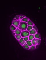
Caenorhabditis elegans is a multicellular organism that has been used to study endocytic trafficking. Originally, assays were developed to study endocytosis in C. elegans oocytes, embryos, and coelomocytes (scavenger cells). Briefly, the assays in oocytes and embryos were performed by measuring the intensities and sizes of compartments containing a yolk protein-green fluorescent protein reporter (VIT-2::GFP) in intestinal compartments at the comma to ‘1.5 fold’ stages of development (Grant and Hirsh, 1999; Schaheen et al., 2006a). In adults, the intensities and sizes of compartments containing GFP (secreted from body wall muscle cells into the psuedocoelom and endocytosed by coelomocytes) were measured in the coelomocytes of transgenic adult C. elegans expressing Pmyo-3::ssGFP (signal sequence-GFP fusion protein) (Treusch et al., 2004). These assays have been used to identify and to elucidate functions of mediators of the endocytic pathway (Fares and Greenwald, 2001a and 2001b; Schaheen et al., 2006b; Huynh et al., 2016).
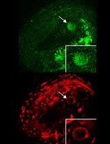
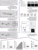
Here, we present an assay that can be used to study endocytosis in another C. elegans tissue. Adult intestinal cells of C. elegans are large and are thus also a great system for deciphering the endocytic pathway using live imaging techniques. The functions of intestinal cells include food assimilation and synthesis, storage of macromolecules, stress response, and host-pathogen interactions (McGhee, 2007). However, one of the main challenges of studying endocytic transport by live imaging in adult intestinal cells is the prevalence of auto-fluorescent gut granules that interfere with the unambiguous determination of bona fide endocytic compartment reporter (like GFP) signals and therefore bias qualitative and quantitative studies (Clokey and Jacobson, 1986). We therefore adapted a method developed by the Grant laboratory to conclusively identify signals from reporters in adult intestinal cells (Gleason et al., 2016; Huynh et al., 2016).
Materials and Reagents
- Microscope slides (VWR, catalog number: 48382-171 )
- Coverslips for microscope slides (Fisher Scientific, Fisherbrand, catalog number: 12-541A )
- 60 mm plates (Fisher Scientific, Fisherbrand, catalog number: FB0875713A )
- 100 mm plates (Fisher Scientific, Fisherbrand, catalog number: FB0875713 )
- Labeling tape (Fisher Scientific, Fisherbrand, catalog number: 15-901-5K )
- Glass pipette
- Aluminum foil
- Autoclave tape
- Inoculating loops
- Pipette tips
- C. elegans experimental strain:
RT258: unc-119(ed3); pwIs50[lmp-1::GFP, unc-119]
Note: LMP-1 is the orthologue of mammalian Lamp1 that localizes to lysosomes in Caenorhabditis elegans (Kostich et al., 2000) - C. elegans control strain: N2
- Calcium chloride dihydrate (CaCl2·2H2O) (Fisher Scientific, catalog number: C79-500 )
- Magnesium sulfate heptahydrate (MgSO4·7H2O) (Fisher Scientific, catalog number: M63-500 )
- Potassium phosphate monobasic (KH2PO4) (Fisher Scientific, catalog number P285-500 )
- Cholesterol (Sigma-Aldrich, catalog number: C8667 )
- EtOH (Merck, catalog number: EX0276-4 )
- LE agarose for making 2.2% agarose (BioExpress, GeneMate, catalog number: E-3120 )
- Levamisole (Sigma-Aldrich, catalog number: 31742 )
- C. elegans culture
- Platinum wire-pick to transfer C. elegans
- Making NGM plates (Brenner, 1974; He, 2011) (see Recipes)
Sodium chloride (NaCl) (Fisher Scientific, catalog number: S271-3 )
Bacto peptone (BD, BactoTM, catalog number: 211677 )
Bacto agar (BD, BactoTM, catalog number: 214030 )
Double distilled water
Cholesterol 5 mg/ml in 95% EtOH (see Recipes)
1 M CaCl2 sterile (see Recipes)
1 M MgSO4 sterile (see Recipes)
1 M KH2PO4 pH 6.0 sterile (see Recipes) - Making 2x YT + OP50 for spotting NGM plates
OP50 frozen stock
2x YT agar plate - 2x YT agar plate (see Recipes)
Bacto tryptone (BD, BactoTM, catalog number: 211705 )
Yeast extract (Fisher Scientific, catalog number: BP1422-500 )
Sodium chloride (NaCl) (Fisher Scientific, catalog number: S271-3 )
Bacto agar (BD, catalog number: 214030 )
Double distilled water - 2x YT medium (see Recipes)
- 2x YT medium + OP50 (see Recipes)
- Platinum wire-pick to transfer C. elegans
- 2.2% agarose pad (see Recipes)
- 9 mM levamisole/1x PBS (see Recipes)
Equipment
- 20 °C Incubator for C. elegans storage (VWR, manufactured by Sheldon Manufacturing, model: Model 2020 )
- 4 L flask
- Stir bar
- Stir plate
- Autoclave
- 2 L beaker
- 1 L bottle
- 37 °C Incubator (VWR, manufactured by Sheldon Manufacturing, model: 5025 T )
- Microwave
- Heating block
- 5-100 ml bottle
- Microscope (Carl Zeiss, model: STEMI SV 6 )
- Zeiss LSM 510 Meta confocal microscope (Zeiss, model: LSM 510 ):
- 63x lens
- Argon 488-nm laser
- Helium neon 543-nm laser
- 63x lens
Software
- MetaMorph® Microscopy Automation & Image Analysis Software (Sunnyvale, CA)
Procedure
文章信息
版权信息
© 2018 The Authors; exclusive licensee Bio-protocol LLC.
如何引用
Huynh, J. M., Dang, H. and Fares, H. (2018). Measurement of Lysosomal Size and Lysosomal Marker Intensities in Adult Caenorhabditis elegans. Bio-protocol 8(3): e2724. DOI: 10.21769/BioProtoc.2724.
分类
细胞生物学 > 细胞成像 > 活细胞成像
您对这篇实验方法有问题吗?
在此处发布您的问题,我们将邀请本文作者来回答。同时,我们会将您的问题发布到Bio-protocol Exchange,以便寻求社区成员的帮助。