

Immunofluorescence Analysis of Human Endocervical Tissue Explants Infected with Neisseria gonorrhoeae
感染淋病奈瑟菌的人宫颈内组织外植体的免疫荧光分析
发布: 2018年02月05日第8卷第3期 DOI: 10.21769/BioProtoc.2720 浏览次数: 7956
评审: Lokesh KalekarBenoit ChassaingAnonymous reviewer(s)
参见作者原研究论文
The authors used this protocol in:

Apr 2017
Advertisement


Abstract
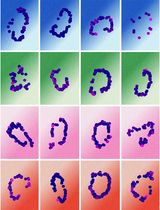
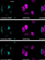
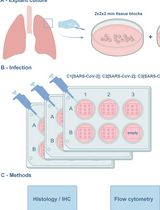
Colonization and penetration of the epithelium is the infection-initiating route of mucosal pathogens. The epithelium counteracts infection by eliciting host cell responses while maintaining the mucosal barrier function. The obligate human sexually transmitted bacterium Neisseria gonorrhoeae, or gonococcus (GC) infects the female reproductive tract primarily from the endocervical epithelium. Due to lack of an infection model that mimics all aspects of human infections in the female reproductive tract, GC pathogenesis is poorly understood. This protocol takes advantage of the viability and functional integrity of human cervical tissues propagated in culture to generate an ex vivo infection model. This tissue model maintains the nature of the infection target and environment without any manipulation such as immortalization of epithelial cells by viruses. Using immunofluorescence microscopy, the interaction of GC with the endocervical epithelium was analyzed.
Keywords: Neisseria gonorrhoeae (淋病奈瑟菌)Background
Neisseria gonorrhoeae (GC) infects human genital epithelium causing gonorrhea, a common sexually transmitted infection. Infections in women can lead to severe complications, such as pelvic inflammatory disease, causing fallopian tube scarring and blockage and predisposition to ectopic pregnancy or infertility. Gonorrhea has reemerged as a critical public health issue due to increased prevalence of antibiotic-resistant strains. Because humans are the only host for GC, a lack of an infection model that mimics all aspects of human infections has been a major obstacle to advance our understanding of GC pathogenesis. We have established a human endocervical tissue explant model and immunofluorescence microscopic analysis to examine the mechanism by which GC infect the human endocervix, the primary site for GC infection in women. This ex vivo model maintains the normal cytoarchitecture and tissue integrity of the endocervical epithelium. Using this model and immunofluorescence analysis, we demonstrate that GC colonizes and penetrates into the endocervical tissue, where they potentially cause symptomatic and disseminated gonococcal infection. GC penetration is enabled by the junction disruption and exfoliation of endocervical epithelial cells in response to GC infection. Taken together, our data show that GC infection in endocervical tissue explants resembles GC infection in vivo observed using patients’ biopsies. In combination with immunofluorescent microscopy, this infection model removes an important roadblock to fully understanding the pathogenesis of GC.
Materials and Reagents
- Cotton swab (Fisher Scientific, catalog number: 23-400-122 )
Manufacturer: Thermo Fisher Scientific, catalog number: 16F0024 . - Carbon steel surgical blades #20 (Aspen Surgical, Bard-Parker, catalog number: 371120 )
- Petri dishes (VWR, catalog number: 25384-302 )
- 6-well tissue culture plates (Corning, Falcon®, catalog number: 353046 )
- Sterile Microcentrifuge tubes (1.7 ml) (Sorenson BioScience, catalog number: 16070 )
- Sterile 15 ml conical tubes (VWR, catalog number: 21008-216 )
- Sterile polyester-tipped applicators (Fisher Scientific, catalog number: 23-400-122 )
- Pipette tips (1,000 μl: VWR, catalog number: 83007-382 ; 200 μl: VWR, catalog number: 53509-007 ; 0.1-10 μl: Fisher Scientific, catalog number: 02-717-133 )
- PAP pen for immunostaining (Sigma-Aldrich, catalog number: Z377821-1EA )
Manufacturer: TED PELLA, catalog number: 22309 . - VWR Micro Slides Superfrost Plus (VWR, catalog number: 48311-703 )
- Zeiss NO.1.5 cover glasses (ZEISS, catalog number: 474030-9000-000 )
- Polysciences flat embedding mold 1.2 x 0.5 x 0.4 cm (Length x Width x Depth) (Polysciences, catalog number: 02615 )
- MS11 Neisseria gonorrhoeae strain(s) (kindly provided by Dr. Herman Schneider, Walter Reed Army Institute for Research)
- Hank’s balanced salt solution (HBSS) (Thermo Fisher Scientific, GibcoTM, catalog number: 14025092 )
- Liquid nitrogen (Roberts Oxygen Company)
- Hoechst 33342, trihydrochloride, trihydrate, 10 mg/ml (Thermo Fisher Scientific, InvitrogenTM, catalog number: H3570 )
- Alexa Fluor 488 goat anti-mouse IgG1 (Thermo Fisher Scientific, Invitrogen, catalog number: A-21121 )
- Mouse anti-human ZO1 (BD, Transduction LaboratoriesTM, catalog number: 610966 )
- Goat anti-GC antibody (Lab generated)
- DyLight 633 Antibody Labeling Kit (Thermo Fisher Scientific, Thermo ScientificTM, catalog number: 53046 )
- Aqua Poly/Mount (Polysciences, catalog number: 18606 )
- Nail polish (Electron Microscopy Science, catalog number: 72180 )
- O.C.T. Compound (SAKURA, Tissue-Tek, catalog number: 4583 )
- Penicillin G sodium salt (Sigma-Aldrich, catalog number: P3032-25MU )
- Streptomycin sulfate (Sigma-Aldrich, catalog number: S9137 )
- Leibovitz’s (1x) L-15 medium (Thermo Fisher Scientific, GibcoTM, catalog number: 21083027 )
- CMRL medium 1066 (1x) (Thermo Fisher Scientific, GibcoTM, catalog number: 11530037 )
- Hydrocortisone 21-hemisuccinate sodium (Sigma-Aldrich, catalog number: H2270-100MG )
- Bovine insulin (Akron Biotech, catalog number: AK8213-0100 )
- L-glutamine 200 mM (100x) (Thermo Fisher Scientific, GibcoTM, catalog number: 25030081 )
- Fetal bovine serum (FBS) (Thermo Fisher Scientific, GibcoTM, catalog number: 10082147 )
- DifcoTM GC medium base (BD, DifcoTM, catalog number: 228950 )
- Agar (United States Biological, catalog number: A0930 )
- L-glutamine, White Crystals or Crystalline Powder (Fisher Scientific, catalog number: BP379-100 )
- Ferric nitrate, nonahydrate (Sigma-Aldrich, catalog number: 254223-10G )
- Thiamine pyrophosphate (Sigma-Aldrich, catalog number: C8754-5G )
- Glucose, as Dextrose anhydrous (Fisher Scientific, catalog number: BP350-1 )
- Sodium chloride (NaCl) (Fisher Scientific, catalog number: S671-10 )
- Potassium chloride (KCl) (Sigma-Aldrich, catalog number: P9333-500G )
- Sodium phosphate dibasic (Na2HPO4) (Fisher Scientific, catalog number: S374-1 )
- Potassium phosphate monobasic (KH2PO4) (Fisher Scientific, catalog number: BP329-1 )
- Hydrochloric acid (HCl) (Fisher Scientific, catalog number: A144-212 )
Manufacturer: Thermo Fisher Scientific, catalog number: FLA144212 . - Paraformaldehyde 16% solution (Electron Microscopy Science, catalog number: 15710 )
- Sucrose (Merck, catalog number: SX1075-1 )
- Gelatin from porcine skin (Sigma-Aldrich, catalog number: G1890-500G )
- Gelatin from cold water fish skin (FSG) (Sigma-Aldrich, catalog number: G7765-250ML )
- Triton X-100 (Sigma-Aldrich, catalog number: X100-100ML )
- 100x penicillin/streptomycin stock (see Recipes)
- Cervical tissue culture medium (see Recipes)
- GCK agar plate (see Recipes)
- 100x Kellogg’s supplement (see Recipes)
- 1x phosphate buffer solution (PBS) (see Recipes)
- 4% paraformaldehyde (PFA) (see Recipes)
- 5% and 15% sucrose solution (see Recipes)
- 7.5% and 20% gelatin solution (see Recipes)
- Staining solution (see Recipes)
Equipment
- Biosafety cabinet (NU-425-600 Class II, A2 Laminar Flow Biohazard Hood) (Nuaire, model: NU-425-600 Class II, A2 , catalog number: 32776)
- Spectrophotometer Ultrospec 2000 UV (Pharmacia Biotech, model number: 80-2106-00 )
- Heating block (Thermo Fisher Scientific, Thermo ScientificTM, catalog number: SP18425Q )
- Stainless steel forceps (VWR, Dissecting forceps)
- Multi-Purpose rotator (Multi-Purpose Rotator) (Barnstead International Lab-line, Thermo Fisher Scientific, Thermo ScientificTM, model: Model 2309 )
- CO2 incubator (Fisher Scientific, model: Isotemp Incubator Model 3530 )
- Confocal microscope (Carl Zeiss, model: LSM 710 )
- Cryostat (Thermo Fisher Scientific, model: Microm HM 550-388114 )
- Slide holder (Newcomer Supply, catalog number: 6841 )
Software
- NIH ImageJ and ZEN software
- GraphPad Software (La Jolla, CA)
Procedure
文章信息
版权信息
© 2018 The Authors; exclusive licensee Bio-protocol LLC.
如何引用
Wang, L., Yu, Q., Stein, D. C. and Song, W. (2018). Immunofluorescence Analysis of Human Endocervical Tissue Explants Infected with Neisseria gonorrhoeae. Bio-protocol 8(3): e2720. DOI: 10.21769/BioProtoc.2720.
分类
微生物学 > 微生物-宿主相互作用 > 离体模型
免疫学 > 粘膜免疫学 > 上皮
细胞生物学 > 细胞成像 > 荧光
您对这篇实验方法有问题吗?
在此处发布您的问题,我们将邀请本文作者来回答。同时,我们会将您的问题发布到Bio-protocol Exchange,以便寻求社区成员的帮助。