

Common Carotid Arteries Occlusion Surgery in Adult Rats as a Model of Chronic Cerebral Hypoperfusion
成年大鼠颈总动脉闭塞手术作为慢性脑低灌注模型

发布: 2018年01月20日第8卷第2期 DOI: 10.21769/BioProtoc.2704 浏览次数: 11936
评审: Oneil G. BhalalaAlessandro DidonnaEdel Hennessy
参见作者原研究论文
The authors used this protocol in:
May 2017


Abstract
Chronic cerebral hypoperfusion (CCH) is an important risk factor of vascular dementia (VaD) and Alzheimer’s disease (AD). Hypoxia/ischemia in the whole brain induced by CCH causes serious damage to brain structure and function, which can lead to cognitive impairment. Two-vessel occlusion (2-VO), also known as permanent, bilateral common carotid artery occlusion, is one of the most widely used animal models (e.g., rat) of CCH to investigate the mechanisms of neurodegenerative processes. In this protocol, we present the surgical procedure for 2-VO in rats.
Keywords: Chronic cerebral hypoperfusion (慢性脑灌注不足)Background
Many neurological and psychiatric illnesses are caused by disorders of the cerebral circulation. A sudden interruption of the blood supply to distinct brain regions can lead to stroke, while a gradual reduction of continuous cerebral blood flow (CBF) impairs memory processes and contributes to the development of dementia (Farkas and Luiten, 2001; Matsuda, 2001; de la Torre, 2002). In the 2-VO rat model, there is a dramatic decrease in CBF to the brain during the acute ischemic phase (2-3 days post-operation) and the cortical and white matter areas have the largest decrease in blood flow, reaching 35%-45% of the control level (Otori et al., 2003). In the chronic ischemic phase (1-3 months post-operation), the CBF values begin to gradually recover at 1 week, but are still significantly lower than the control values 4 weeks after 2-VO induction (Schmidt-Kastner et al., 2001; Otori et al., 2003; Tomimoto et al., 2003). After 8 weeks to 3 months of 2-VO, only a slight reduction or virtually no reduction of flow has been reported (Otori et al., 2003). Finally, after 6 months of 2-VO, the CBF almost returns completely to normal (Choy et al., 2006), because other arterial sources of blood provide compensatory blood flow (via the circle of Willis, Figure 1) to areas that typically are supplied by the common carotid (Farkas et al., 2007). The 2-VO model exhibits characteristic features of human CCH condition, such as cerebral blood flow and metabolic changes (Ohta et al., 1997; Otori et al., 2003), learning and memory disturbances (Farkas and Luiten, 2001; Farkas et al., 2004b; Liu et al., 2005) and the neuropathologic changes (Kreutzberg, 1996; Farkas et al., 2004b; Panickar and Norenberg, 2005; Ohtaki et al., 2006; Eisel et al., 2006). Additionally, this model has been used to study cerebrovascular WM lesions (Wakita et al., 2002; Takizawa et al., 2003; Farkas et al., 2004a).
In the traditional 2-VO experiment, silk suture is used to ligate the bilateral common carotid artery, and an acute phase after the occlusion with dramatic CBF fall ensues. To improve the 2-VO model, researchers have tested some alternative methods. For example, a silicone collar cuff can be placed around the common carotid artery in order to reproduce the inflammatory response caused by atherosclerosis. However, this operation does not cause long-term memory impairment (de Bortoli et al., 2005). A study in which the two common carotid arteries were occluded at intervals of 1 week found that procedure leads to a progressive decrease in brain perfusion, and decreased mortality compared to procedures that occlude both arteries at once (Sarti et al., 2002a and 2002b). But an undesirable feature of these protocols is that the rats must undergo anesthesia twice a week, which can be stressful to the animal. Kitaguchia et al. (2009) use a 30 min delay between carotid arteries in the murine BCAS model to decrease mortality. Other research groups use ameroid constrictors in place of the silk suture. The ameroid constrictors consist of a titanium shell surrounding the hygroscopic casein material with the internal lumen. The casein component gradually absorbs water and thus expands, resulting in narrowing and occlusion of its encased arterial lumen. So the reduction of the CBF could be mild without a sharp drop (Hattori et al., 2015). Here, we described a protocol in detail for performing 2-VO in rat.
Figure 1. Schematic diagram of the circle of Willis in human (A) and rat (B). ACA (anterior cerebral artery); ACOA (anterior communicating artery); AICA (anterior inferior cerebellar artery); ASA (anterior spinal artery); BA (basilar artery); ICA (internal carotid artery); MCA (middle cerebral artery); PCA (posterior cerebral artery); PCOA (posterior communicating artery); SCA (superior cerebellar artery); VA (vertebral artery). This figure is adapted from Eszter Farkas et al. (2001).
Materials and Reagents
- Cotton balls (Beijing Sunny Medical Technology Development, catalog number: YG-048 )
- Cotton swabs (Beijing Sunny Medical Technology Development, catalog number: YG-053 )
- Silk suture (3/0) (Shanghai Pudong Jinhuan Medical Supplies, 1#)
- Male adult Sprague Dawley (SD)/Wistar rats at 10-12 weeks of age (or based on experimental needs to determine the age of rats) (Beijing Vital River Laboratory Animal Technology)
- 75% ethanol (ANNJET, Q/371402AAJ008)
- Iodine tincture (ANNJET, Q/371402AAJ001)
- 10% chloral hydrate (300 mg/kg intraperitoneally, Sinopharm Chemical Reagent, catalog number: 30037516 )
- 0.9% sodium chloride solution (Shijiazhuang No.4 Pharmaceutical, H13023201)
- 10% chloral hydrate (see Recipes)
Equipment
- Rat dissection board (BEIJING HELI KECHUANG TECHNOLOGY DEVELOPMENT, model: HL/JPT-2 )
- Electronic balance (Shanghai Yoke Instrument, catalog number: YP10002 )
- Curved ophthalmic scissors (Shanghai Medical Instruments, catalog number: Y00020 ) (Figure 2D)
- Medical suture needle (HANGZHOU HUAWEI MEDICAL APPLIANCE, catalog number: P1531 ) (www.hzhwyl.com.cn)
- Operating scissors (Shanghai Medical Instruments, catalog number: J21010 ) (Figure 2B)
- Straight ophthalmic scissors (Shanghai Medical Instruments, catalog number: Y00030 ) (Figure 2C)
- Hemostatic forceps (Shanghai Medical Instruments, catalog number: J31050 ) (Figure 2E)
- Ophthalmic forceps (Shanghai Medical Instruments, catalog number: JD1020 ) (Figure 2F)
- Tissue holding forceps (Shanghai Medical Instruments, catalog number: JD2010 ) (Figure 2G)
- Needle holder (Shanghai Medical Instruments, catalog number: J32020 ) (Figure 2A)
- Sterilizing trays (Shanghai Medical Instruments, catalog number: R0B030 )
- Syringe with needle (Shanghai Zhiyu Medical Equipment, 2 ml)
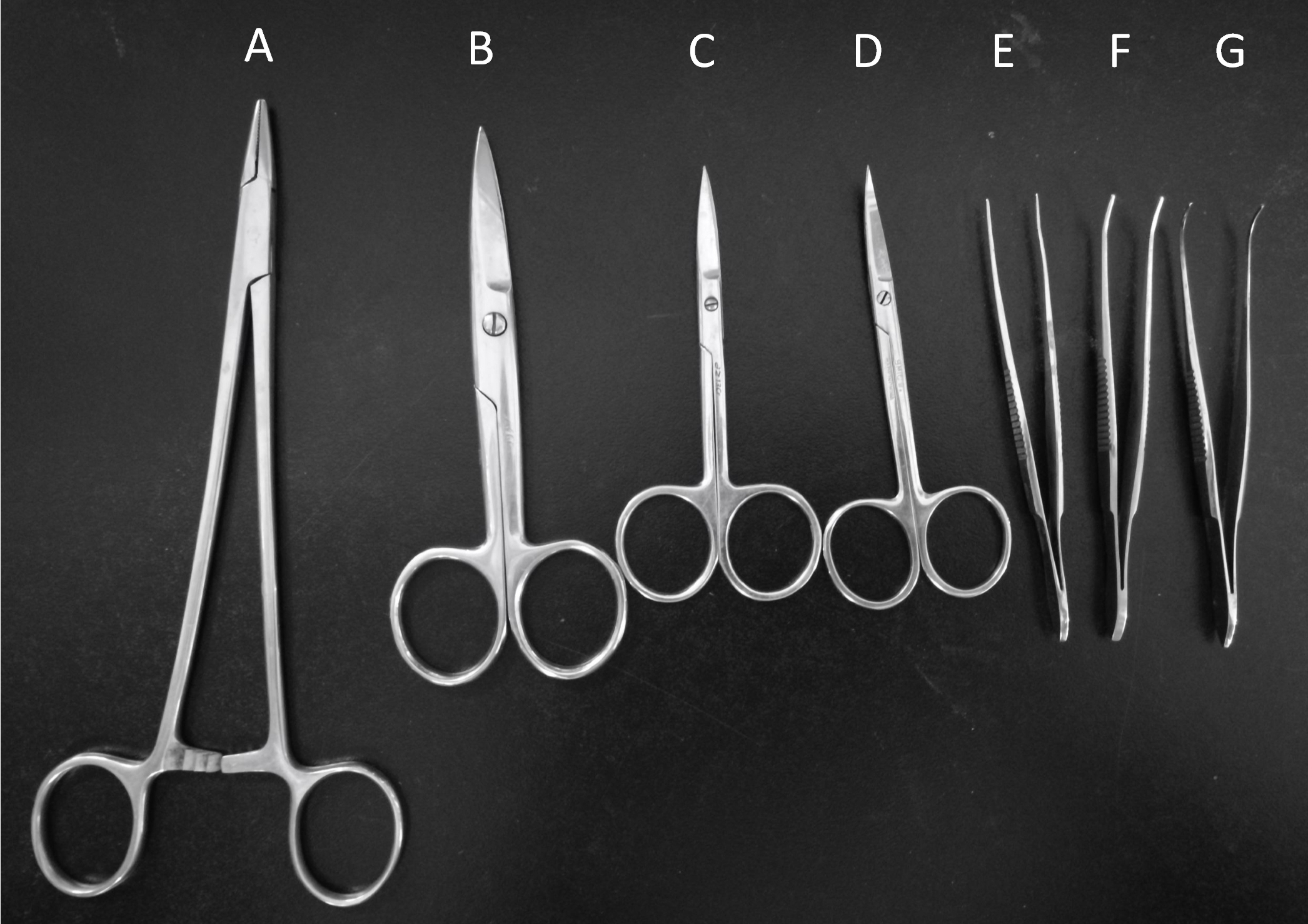
Figure 2. Surgery tools. Needle holder (A), Operating scissors (B), Straight ophthalmic scissors (C), Curved ophthalmic scissors (D), Hemostatic forceps (E), Ophthalmic forceps (F), Tissue holding forceps (G).
Procedure
文章信息
版权信息
© 2018 The Authors; exclusive licensee Bio-protocol LLC.
如何引用
Cao, D., Bai, Y. and Li, L. (2018). Common Carotid Arteries Occlusion Surgery in Adult Rats as a Model of Chronic Cerebral Hypoperfusion. Bio-protocol 8(2): e2704. DOI: 10.21769/BioProtoc.2704.
分类
神经科学 > 神经系统疾病 > 动物模型
分子生物学 > 蛋白质 > 表达
细胞生物学 > 组织分析 > 生理学
您对这篇实验方法有问题吗?
在此处发布您的问题,我们将邀请本文作者来回答。同时,我们会将您的问题发布到Bio-protocol Exchange,以便寻求社区成员的帮助。