

Live Imaging of Axonal Transport in the Motor Neurons of Drosophila Larvae
果蝇幼虫运动神经元中轴突运输的实时成像
 2.jpg)
发布: 2017年12月05日第7卷第23期 DOI: 10.21769/BioProtoc.2631 浏览次数: 9860
评审: Jihyun KimAdler R. DillmanAnonymous reviewer(s)
参见作者原研究论文
The authors used this protocol in:
Jul 2017
Advertisement


Abstract
Axonal transport, which is composed of microtubules, motor proteins and a variety of types of cargo, is a prominent feature of neurons. Monitoring these molecular dynamics is important to understand the biological processes of neurons as well as neurodegenerative disorders that are associated with axonal dysfunction. Here, we describe a protocol for monitoring the axonal transport of motor neurons in Drosophila larvae using inverted fluorescence microscopy.
Keywords: Axonal transport (轴突运输)Background
Axons are a unique neuronal structure, by which neurons transmit electric and chemical signals to neighboring neurons, muscles and other tissues. Axonal function is maintained by the circular flow or shuttling of vesicles, organelles and materials (Liu et al., 2012; Wong et al., 2012; Alami et al., 2014). Axonal dysfunction is thought to be an early sign of neurodegeneration and a cause of neurodegenerative diseases such as Alzheimer’s, Parkinson’s and motor neuron diseases in humans (Hirokawa et al., 2010; Millecamps and Julien, 2013). However, it is difficult to monitor the process of axonal degeneration in these neurodegenerative diseases in both humans and mammalian models. The Drosophila model is a powerful tool for studying neurodegenerative diseases at the molecular-genetic level and has provided important evidence for therapeutic approaches through the live imaging of neurons in vivo (Shiba-Fukushima et al., 2014; Hosaka et al., 2017). Upright fluorescence microscopy techniques are generally used to analyze the live imaging of tissues and organ cultures. However, inverted fluorescence microscopy has seen an increase in demand due to its broad utility and extensibility. Here, we introduce our protocol to analyze the axonal transport of motor neurons in the third-instar larvae of Drosophila using inverted fluorescence microscopy.
Materials and Reagents
- 35-mm culture dishes (Thermo Fisher Scientific, Thermo ScientificTM, catalog number: 153066 )
- Single sided plastic tape (0.2 mm in thickness) (3M, catalog number: 35 )
- Eight insect pins (Fine Science Tools, catalog number: 26002-10 , Minutien pins, tip diameter 0.0125 mm, rod diameter 0.1 mm, stainless steel)
Note: The heads of 6 pins are shortened to approximately 3 mm in length for dissection (Figures 1 and 4); the other 2 are not modified and are used for the observation tank (Figures 1 and 3).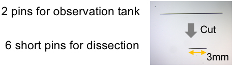
Figure 1. Preparation of pins - Silicone (Shin-Etsu Silicone, KE-106 and CAT-RG)
- Cover glasses (24 x 40 mm and 18 x 18 mm, 0.12-0.17 mm in thickness, Matsunami Glass, catalog numbers: C024401 and C218181 )
- Drosophila melanogaster-harboring gene mutations or transgenes (most strains are available from public stock centers, which can be accessed through the website below: http://flybase.org/wiki/FlyBase:Stocks)
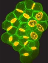
Note: We utilized the following lines in Figure 6 and our previous study (Hosaka et al., 2017): UAS-preproANF-EMD; D42-GAL4 (Bloomington Drosophila Stock Center, catalog numbers: 7001 and 8816). This line expresses preproANF-EMD, which visualizes the dense core vesicles in motor neurons with green fluorescence. - Sodium chloride (NaCl) (NACALAI TESQUE, catalog number: 31319-45 )
- Potassium chloride (KCl) (Wako Pure Chemical Industries, catalog number: 163-03545 )
- Magnesium chloride hexahydrate (MgCl2·6H2O) (Wako Pure Chemical Industries, catalog number: 135-00165 )
- Sodium bicarbonate (NaHCO3) (Sigma-Aldrich, catalog number: S6014 )
- Sucrose (Wako Pure Chemical Industries, catalog number: 196-00015 )
- Trehalose (Sigma-Aldrich, catalog number: T0167 )
- HEPES (Sigma-Aldrich, catalog number: H4034 )
- Calcium chloride dihydrate (CaCl2·2H2O) (NACALAI TESQUE, catalog number: 06731-05 )
- Modified HL-3 solution (Stewart et al., 1994) (see Recipes)
Equipment
- Tweezers (Fine Science Tools, catalog number: 11251-30 , Dumont #5 forceps dumoxel standard tip)
- Micro-scissors (World Precision Instruments, catalog number: 501778 , SuperFine vannas scissors, 3 mm straight blades)
- Incubator (Panasonic Healthcare, catalog number: MIR-262 )
- Inverted fluorescence microscopy (Leica Microsystems, model: Leica TCS SP5 )
Software
- ImageJ software (National Institutes of Health) for image analysis
Procedure
文章信息
版权信息
© 2017 The Authors; exclusive licensee Bio-protocol LLC.
如何引用
Inoshita, T., Hattori, N. and Imai, Y. (2017). Live Imaging of Axonal Transport in the Motor Neurons of Drosophila Larvae. Bio-protocol 7(23): e2631. DOI: 10.21769/BioProtoc.2631.
分类
神经科学 > 神经系统疾病 > 动物模型
神经科学 > 细胞机理 > 胞内信号传导
细胞生物学 > 细胞成像 > 活细胞成像
您对这篇实验方法有问题吗?
在此处发布您的问题,我们将邀请本文作者来回答。同时,我们会将您的问题发布到Bio-protocol Exchange,以便寻求社区成员的帮助。