

Obtaining Multi-electrode Array Recordings from Human Induced Pluripotent Stem Cell–Derived Neurons
从人诱导性多能干细胞神经元获得多电极阵列记录
发布: 2017年11月20日第7卷第22期 DOI: 10.21769/BioProtoc.2609 浏览次数: 13119
评审: Jihyun KimKhyati Hitesh ShahAnonymous reviewer(s)
参见作者原研究论文
The authors used this protocol in:
Mar 2017


Abstract
Neuronal electrical properties are often aberrant in neurological disorders. Human induced pluripotent stem cells (hiPSCs)-derived neurons represent a useful platform for neurological disease modeling, drug discovery and toxicity screening in vitro. Multi-electrode array (MEA) systems offer a non-invasive and label-free platform to record neuronal evoked-responses concurrently from multiple electrodes. To better detect the neural network changes, we used the Axion Maestro MEA platform to assess neuronal activity and bursting behaviors in hiPSC-derived neuronal cultures. Here we describe the detailed protocol for neuronal culture preparation, MEA recording, and data analysis, which we hope will benefit other researchers in the field.
Keywords: Multi-electrode array (多电极阵列)Background
Human induced pluripotent stem cell (hiPSC) technology is currently being used to model neurological and psychiatric diseases in vitro. Recent studies have demonstrated that certain cellular phenotypes associated with particular disorders can be recapitulated in the dish. Neural electrical activity is at the essence of nervous system function, representing a key form of communication whose normal functioning is essential for emotion, memory, sensory modalities, and behavior in vivo. In disease conditions, the electrical properties can be affected, so it is important to understand neuronal circuit connectivity, physiology, and pathology in hiPSC-based models of neurological disease.
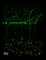
Patch-clamp and multi-electrode array (MEA) technology are the prevailing techniques used to assess electrophysiological activity, and thus neuronal function. While patch-clamp is a powerful intracellular method to investigate the activity and function of a single cell (Neher et al., 1978), an MEA plate has the ability to record extracellular action potentials (or spikes) and local field potentials simultaneously from thousands of different cells in the same plate over time, thus offering a better understanding of neuronal activity at a network level (Hutzler et al., 2006; Obien et al., 2014). MEAs are grids of tightly spaced electrodes that are capable of directly sensing changes in extracellular membrane potential in excitable cells and produce real-time trace of neuronal activity. The transparent CytoView 12-well MEA plate contains 64 recording electrodes per well, whereas the 48-well MEA plate contains 12 electrodes per well. The MEA system then parses these voltage traces for action potential waveforms, time-stamps the waveforms, and calculates action potential firing rates on-line for each electrode. This on-line firing rate is represented by color-coded heat-maps that depict real-time and simultaneous indications of network activity, and experimental effects (Figure 1).
Figure 1. Example plate activity heat map, and spike detector. Top: Heat map of MEA reading visualized using Axion BioSystems Integrated Studio (AxIS). Each box represents one well. Active channels represented by bright light blue dots on the map. Bottom: Offline spiking activity illustrated on AxIS data display.
Materials and Reagents
- 6-well cell culture plate (Corning, Costar®, catalog number: 3516 )
- Plastic pipette tips (Corning, Axygen®, catalog number: T-1000-B )
- Sterile 1.5 ml centrifuge tubes (Corning, Axygen®, catalog number: MCT-175-C )
- 15 ml and 50 ml centrifuge tubes (Corning, Falcon®, catalog numbers: 352096 and 352070 )
- Petri dish (Corning, Falcon®, catalog number: 351029 )
- Cell lifter (Corning, catalog number: 3008 )
- 12-well MEA plate (Axion BioSystem, catalog number: M768-GL1-30Pt200 )
- 0.22 µm filter (Corning, catalog number: 431097 )
- Kimwipes (KCWW, Kimberly-Clark, catalog number: 34155 )
- Sterile water (Thermo Fisher Scientific, GibcoTM, catalog number: 15230162 )
- Dispase (STEMCELL Technologies, catalog number: 07923 )
- Y-27632 (STEMCELL Technologies, catalog number: 72302 )
- Laminin (Thermo Fisher Scientific, GibcoTM, catalog number: 23017015 )
- LDN-193819 (Stemgent, catalog number: 04-0074 )
- SB431542 (Sigma-Aldrich, catalog number: S4317 )
- XAV939 (Stemgent, catalog number: 04-0046 )
- Recombinant Human Sonic Hedgehog (SHH) (R&D Systems, catalog number: 464-SH-025 )
- Brain-derived neurotrophic factor (BDNF) (R&D Systems, catalog number: 248-BD-025 )
- Glial-derived neurotrophic factor (GDNF) (R&D Systems, catalog number: 212-GD-050 )
- L-Ascorbic acid (Sigma-Aldrich, catalog number: A4403 )
- N6,2’-O-Dibutyryladenosine 3’,5’-cyclic monophosphate sodium salt (dbcAMP) (Sigma-Aldrich, catalog number: D0260 )
- 50% poly(ethyleneimine) (PEI) solution (Sigma-Aldrich, catalog number: P3143 )
- Accutase (STEMCELL Technologies, catalog number: 07920 )
- Tetrodotoxin (TTX) (Alomone Labs, catalog number: T-500 )
- N2 (Thermo Fisher Scientific, GibcoTM, catalog number: 17502048 )
- B27 (Thermo Fisher Scientific, GibcoTM, catalog number: 17504044 )
- Modified Eagle’s Medium-Non Essential Amino Acid (MEM-NEAA) (Thermo Fisher Scientific, GibcoTM, catalog number: 11140050 )
- L-Glutamine (Thermo Fisher Scientific, GibcoTM, catalog number: 25030081 )
- Penicillin-streptomycin (Pen-Strep) (Thermo Fisher Scientific, GibcoTM, catalog number: 15140122 )
- Dulbecco’s modified Eagle’s medium: Nutrient Mixture F-12 (DMEM/F12) (Thermo Fisher Scientific, GibcoTM, catalog number: 11320082 )
- Neurobasal medium (Thermo Fisher Scientific, GibcoTM, catalog number: 21103049 )
- N2 Supplement-A (STEMCELL Technologies, catalog number: 07152 )
- NeuroCultTM SM1 (STEMCELL Technologies, catalog number: 05711 )
- BrainPhysTM neuronal medium (STEMCELL Technologies, catalog number: 05790 )
- Boric acid (Sigma-Aldrich, catalog number: B6768 )
- Sodium tetraborate (Sigma-Aldrich, catalog number: 221732 )
- Hydrochloric acid fuming 37% (Merck, catalog number: 100317 )
- N2B27 medium (see Recipe 1)
- BrainPhys medium (see Recipe 2)
- Borate buffer (see Recipe 3)
Equipment
- Pipettes
P10 (Gilson, catalog number: F144802 )
P200 (Gilson, catalog number: F123601 )
P1000 (Gilson, catalog number: F123602 ) - Tissue culture hood (Gelman, model: BH Class II Type A2 series )
- 37 °C water bath (Thermo Fisher Scientific, Thermo ScientificTM, model: Labline 183 , catalog number: 2835)
- Cell culture incubator (Thermo Fisher Scientific, Thermo ScientificTM, model: HeracellTM I50i CO2 , catalog number: 50116047)
- Centrifuge (Eppendorf, model: 5810 , catalog number: 5810000424)
- Cell counter Countess II (Thermo Fisher Scientific, model: CountessTM II, catalog number: AMQAX1000 )
- Phase contrast microscope (Leica DMIL LED Inverted Fluorescence microscope) (Leica Microsystems, model: Leica DM IL LED )
- Maestro MEA system (Axion Biosystem)
Software
- Axion Biosystems Integrated Studio (AxIS, Axion Biosystem)
- Neural Metric tool (Axion Biosystem)
- Neuroexplorer (NEX, Plexon)
- MATLAB (R2016a)
- Excel (Microsoft Office)
Procedure
文章信息
版权信息
© 2017 The Authors; exclusive licensee Bio-protocol LLC.
如何引用
Xu, X., Radulescu, C. I., Utami, K. H. and Pouladi, M. A. (2017). Obtaining Multi-electrode Array Recordings from Human Induced Pluripotent Stem Cell–Derived Neurons. Bio-protocol 7(22): e2609. DOI: 10.21769/BioProtoc.2609.
分类
神经科学 > 神经系统疾病 > 细胞机制
干细胞 > 多能干细胞 > 基于细胞的分析方法
神经科学 > 细胞机理 > 细胞分离和培养
您对这篇实验方法有问题吗?
在此处发布您的问题,我们将邀请本文作者来回答。同时,我们会将您的问题发布到Bio-protocol Exchange,以便寻求社区成员的帮助。