

Scanning Electron Microscope (SEM) Imaging to Determine Inflorescence Initiation and Development in Olive
扫描电子显微镜(SEM)观测橄榄花序的起始和发育
.jpg)
发布: 2017年10月05日第7卷第19期 DOI: 10.21769/BioProtoc.2575 浏览次数: 8990
评审: Scott A M McAdamAnonymous reviewer(s)
参见作者原研究论文
The authors used this protocol in:

Jan 2017
Advertisement


Abstract
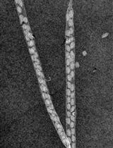
Here we present a protocol that describes how to image the structure of the olive axillary bud meristem with a scanning electron microscope (SEM) in order to characterize its identity and developmental stage. Briefly, the specimen is fixed with glutaraldehyde, saturated with ethanol, dried in a critical point dryer (CPD) system, dissected, coated with a conducting material and imaged with a scanning electron microscopy (SEM).
Keywords: SEM (SEM)Background
The exact timing of flowering induction and inflorescence initiation in olive (Olea europaea L.) is in controversy (Haberman et al., 2017). In olive, inflorescences emerge from lateral buds at the end of winter and flower in the spring. We have developed a protocol to better characterize the timing of inflorescence initiation in olive by imaging the meristem in the olive bud with a SEM at different times during the year. In these SEM images the meristem structure can be identified unambiguously, and the definition level of the meristem can be much higher than images of bud meristem sections presented in previous studies.
Materials and Reagents
- Scalpel blade No. 11 (Sigma-Aldrich, catalog number: S2771 )
- Double-sided adhesive tape
- Glass scintillation vials with screw caps, volume 20 ml (Sigma-Aldrich, catalog number: Z190535 )
- Pipette (BRAND, catalog number: 747760 ) or a similar instrument
- Gold annular target for the sputter coater (Agar scientific, catalog number: AGB7370 )
- Olive (Olea europaea L.) buds
- Sodium phosphate dibasic (Na2HPO4) (Sigma-Aldrich, catalog number: S3264 )
- Sodium phosphate monobasic (NaH2PO4) (Sigma-Aldrich, catalog number: S3139 )
- 25% glutaraldehyde (Sigma-Aldrich, catalog number: G5882 )
- Ethanol absolute (Sigma-Aldrich, catalog number: 24102 )
- Optional: Technical grade ethanol (Sigma-Aldrich, catalog number: V0T0042 )
- 0.1 M phosphate buffer pH 7.2 (sodium phosphate buffer; see Recipes)
- 5% glutaraldehyde solution (in 0.1 M phosphate buffer pH 7.2; see Recipes)
Equipment
- Scalpel handle No. 3 (Sigma-Aldrich, catalog number: S2896 )
- Tweezers style #5 (Sigma-Aldrich, catalog number: T4537 )
- Critical point dryer system (BAL-TEC, model: CPD 030 )
- Stereo-microscope (Olympus, model: SZX12 )
- Sputter coater (E510 scanning electron microscope coating unit) (Polaron Instruments, model: E510 )
- Scanning electron microscope (JEOL, model: JSM-5410 LV )
Procedure
文章信息
版权信息
© 2017 The Authors; exclusive licensee Bio-protocol LLC.
如何引用
Haberman, A., Zelinger, E. and Samach, A. (2017). Scanning Electron Microscope (SEM) Imaging to Determine Inflorescence Initiation and Development in Olive. Bio-protocol 7(19): e2575. DOI: 10.21769/BioProtoc.2575.
分类
植物科学 > 植物发育生物学 > 形态建成
您对这篇实验方法有问题吗?
在此处发布您的问题,我们将邀请本文作者来回答。同时,我们会将您的问题发布到Bio-protocol Exchange,以便寻求社区成员的帮助。