

Staining of Membrane Receptors with Fluorescently-labeled DNA Aptamers for Super-resolution Imaging
用荧光标记的DNA吸附剂染色膜受体以实现超分辨率成像

发布: 2017年09月05日第7卷第17期 DOI: 10.21769/BioProtoc.2541 浏览次数: 10128
评审: Gal HaimovichShalini Low-NamAnca Flavia Savulescu
参见作者原研究论文
The authors used this protocol in:

Feb 2017


Abstract
One of the most prominent applications of fluorescent super-resolution microscopy is the study of nanodomain arrangements of receptors and the endocytic pathway. Staining methods are becoming crucial for answering questions on the nanoscale, therefore, the use of small and monovalent affinity probes is of great interest in super-resolution microscopy with biological samples. One kind of affinity probe is the aptamer. Aptamers are single DNA or RNA sequences that bind with high affinity to their targets and due to their small size they are able to (i) place the fluorophore in close proximity to the protein of interest and (ii) bind to most of the protein of interest overcoming the steric hindrance effect, resulting in better staining density. Here we describe a detailed protocol with which to stain live cells using aptamers and to image them with Stimulated Emission Depletion (STED) microscopy. In this protocol, the stainings were performed with commercially available aptamers that target the epidermal growth factor receptor (EGFR), the human epidermal growth factor receptor 2 (HER2 or ErbB2) and the ephrin type-A receptor 2 (Epha2). Since aptamers can be coupled to most of the popular fluorophores, we believe that the procedure presented here can be extended to the large majority of the current super-resolution microscopy techniques.
Keywords: Microscopy (显微镜)Background
Recent advances in super-resolution imaging techniques have led to the search for more accurate methodologies to tag cellular elements. Diffraction unlimited imaging instruments provide excellent resolutions, however standard sample staining methodologies, such as immunostaining, lack the necessary precision for the detection of cellular elements. Due to their large dimension (~15 nm in length) and high molecular weight (~150 kDa), antibodies can poorly penetrate into biological samples. Additionally, the primary/secondary antibody complex places the fluorophores at approximately 25 nm away from the target, compromising the detection accuracy. Moreover, due to the large size of the primary/secondary antibody complex, a smaller fraction of the targets can be labelled due to the steric hindrance (Fornasiero and Opazo, 2015). This leads to lower labelling density, a crucial parameter for super-resolution microscopy, especially in recognizing and describing nanostructures. To circumvent these problems, small affinity probes that bind to single targets (monovalently) such as aptamers, affibodies or nanobodies have been tested in recent years (Rothbauer et al., 2006; Opazo et al., 2012; Ries et al., 2012) and are becoming valuable tools for super-resolution microscopy. Nanobodies, affibodies and aptamers have a relatively small linear size (~3 nm, ~2 nm and ~3 nm, respectively). This property allows them to place the fluorescent dye in close proximity to the target, penetrate samples in a more efficient manner and bind to a higher fraction of target proteins, bypassing the effect of steric hindrance observed by antibodies (Ries et al., 2012; Mikhaylova et al., 2015). In a previous study we made a systematic comparison between three commercially available aptamers and antibodies that target the epidermal growth factor receptor (EGFR), the human epidermal growth factor receptor 2 (HER2 or ErbB2) and the ephrin type-A receptor 2 (Epha2). Our results showed that aptamers were able to find more epitopes (resulting in higher labeling density). As a consequence, several structural features of the subcellular components that were imaged became more apparent. Among these, the inner lumen and the complex morphology of endocytic organelles were visible. For these reasons, smaller imaging tools are becoming the preferred choice over antibodies, in order to improve the quality of an immunolabeling super-resolution approach and allow a more precise description the localization and the distribution of membrane receptors (Gomes de Castro et al., 2017).
Materials and Reagents
- Biospin 6 column (Micro Bio-SpinTM P-6 Gel Columns, Tris Buffer) (Bio-Rad Laboratories, catalog number: 7326222 )
- Cell culture 12-well plates (Thermo Fisher Scientific, catalog number: 150628 )
- 18 mm diameter round glass coverslips (Gerhard Menzel, catalog number: CB00180RA1 )
- 15 ml tubes
- PCR tubes
- Parafilm M
- Soft paper tissue
- Gloves
- Microscopy glass slides
- 2 ml tubes
- Vacuum filtration (e.g., VWR® Vacuum Filtration Systems, Standard Line) (VWR, catalog number: 10040-436 )
- 0.2 µm syringe filter
- Transfer pipette
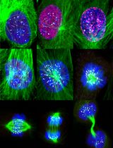
- The images shown in Figure 1 are from aptamers against human:
EGFR (5’-SH-EGFR aptamer-3’, seq # 2369-27-02, 50 mer)
ErbB2 (5’-SH-ErbB2 aptamer-3’, seq # 1194 ± 35, 40 mer)
Epha2 (5’-SH-EphA2 aptamer-3’, seq # 2176-01-01, 76 mer)
Note: They were produced by Aptamer Sciences, Inc., South Korea and supplied by AMS Biotechnology, Europe. All three aptamers contain the chemical modification 5-(N-benzylcarboxyamide)-2’-deoxyuridine (5-BzdU) in unrevealed locations. - Triethylammonium acetate buffer pH 7.0, 1 M (TEAA) (AppliChem, catalog number: A3846 )
- Maleimide Atto647N dye (Atto-TEC, catalog number: AD 647N-41 )
- Dimethyl sulfoxide, anhydrous (DMSO) (Sigma-Aldrich, catalog number: 276855 )
- Sodium chloride (NaCl)
- Ethanol
- 1x Dulbecco’s phosphate buffered saline (1x DPBS) (Sigma-Aldrich, catalog number: D8662 )
- Acetonitrile
- Trypsin-EDTA solution (Lonza, catalog number: 17-161E )
- Ultrapure DNase- and RNase-free distilled water (Carl Roth, catalog number: T143.2 )
- Tris (2-carboxyethyl) phosphine hydrochloride (TCEP) (Sigma-Aldrich, catalog number: C4706 )
- Sodium hydroxide (NaOH)
- DMEM, high glucose medium, no glutamine (Thermo Fisher Scientific, GibcoTM, catalog number: 11960044 )
- Fetal bovine serum (FBS) (Biochrom, catalog number: S 0615 )
- L-Glutamine 200 mM (Lonza, catalog number: BE17-605E )
- Penicillin/streptomycin 10,000 U/ml, each (Lonza, catalog number: 17-602E )
- RPMI 1640 medium, no glutamine (Thermo Fisher Scientific, GibcoTM, catalog number: 21870076 )
- Poly-L-lysine (PLL) (Sigma-Aldrich, catalog number: P5899-5MG )
- Sodium phosphate dibasic (Na2HPO4)
- Potassium phosphate monobasic (KH2PO4)
- Potassium chloride (KCl)
- Magnesium chloride hexahydrate (MgCl2·6H2O)
- Salmon sperm DNA, sheared, 10 mg/ml (Thermo Fisher Scientific, catalog number: AM9680 )
- Dextran sulfate sodium salt (Sigma-Aldrich, catalog number: 31404 )
- Paraformaldehyde (PFA) (Sigma-Aldrich, catalog number: P6148 )
- Glycine (Sigma-Aldrich, catalog number: G8898 )
- Mowiol® (Sigma-Aldrich, catalog number: 81381 )
- 1 M Tris (2-carboxyethyl) phosphine hydrochloride stock solution (TCEP) (see Recipes)
- Complete DMEM medium (see Recipes)
- Complete RPMI medium (see Recipes)
- Poly-L-lysine (PLL) stock solution (see Recipes)
- 5x phosphate buffer saline (5x PBS) (see Recipes)
- 25 mM MgCl2 solution (5x MgCl2) (see Recipes)
- Blocking solution (see Recipes)
- 4% paraformaldehyde (PFA) (see Recipes)
- Quenching solution (see Recipes)
- Mowiol® (see Recipes)
- (Optional) Buffer A (see Recipes)
- (Optional) Buffer B (see Recipes)
Equipment
- Microcentrifuge (e.g., Eppendorf, model: 5415 or similar)
- Nucleosil 100-5 C18 column
- (Optional) Dionex DNAPac PA200 4 x 250 mm column
- Hemocytometer
- Cell culture hood
- Thermal cycler
- Aluminum metal plate (length x width x thickness [cm]: e.g., 20 x 12 x 2)
- Cell culture incubator
- Half-curved-forceps
- Oven
- STED microscope, Leica pulsed STED setup composed by a True Confocal System (TCS) STED SP5 (Leica Microsystems, model: Leica TCS SP5 ) fluorescence microscope equipped with a 100x 1.4 NA HCX PL APO oil objective (Leica Microsystems, Germany)
- Pulsed laser (PicoQuant, Germany)
- Sapphire tunable laser (Mai Tai Broadband, Spectra-Physics, USA)
- Glass beaker
- Magnetic stirrer
- Lab coat, eye protection
Software
- ImageJ (http://imagej.nih.gov/ij/docs/index.html)
- MATLAB (MathWorks, Massachusetts, USA)
Procedure
文章信息
版权信息
© 2017 The Authors; exclusive licensee Bio-protocol LLC.
如何引用
Gomes de Castro, M. A., Höbartner, C. and Opazo, F. (2017). Staining of Membrane Receptors with Fluorescently-labeled DNA Aptamers for Super-resolution Imaging. Bio-protocol 7(17): e2541. DOI: 10.21769/BioProtoc.2541.
分类
细胞生物学 > 细胞成像 > 固定细胞成像
细胞生物学 > 细胞成像 > 荧光
您对这篇实验方法有问题吗?
在此处发布您的问题,我们将邀请本文作者来回答。同时,我们会将您的问题发布到Bio-protocol Exchange,以便寻求社区成员的帮助。