

FM1-43 Photoconversion and Electron Microscopy Analysis at the Drosophila Neuromuscular Junction
果蝇神经肌肉接头处FM1-43光转换和电子显微镜分析

发布: 2017年09月05日第7卷第17期 DOI: 10.21769/BioProtoc.2523 浏览次数: 9165
评审: Pengpeng LiAnonymous reviewer(s)
参见作者原研究论文
The authors used this protocol in:
Jan 2017
Advertisement


Abstract
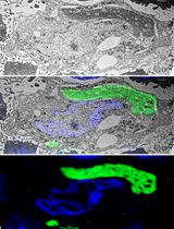
We developed a protocol for photoconversion of endocytic marker FM1-43 followed by electron microscopy analysis of synaptic boutons at the Drosophila neuromuscular junction. This protocol allows detection of stained synaptic vesicle even when release rates are very low, such as during the spontaneous release mode. The preparations are loaded with the FM1-43 dye, pre-fixed, treated and illuminated to photoconvert the dye, and then processed for conventional electron microscopy. This procedure enables clear identification of stained synaptic vesicles at electron micrographs.
Keywords: Electron microscopy (电子显微镜检查)Background
Neuronal transmitters are released via the fusion of synaptic vesicles with the neuronal plasma membrane. Vesicles can fuse spontaneously or in response to an action potential. Subsequently, vesicles become retrieved via endocytosis and recycled. Molecular mechanisms of synaptic vesicle recycling were investigated extensively with the tools of molecular biology, electrophysiology and microscopy (Slepnev and De Camilli, 2000; Sudhof, 2004; Rizzoli and Betz, 2005; Kavalali, 2006). Loading the endocytic marker FM1-43 coupled with the dye photoconversion followed by electron microscopy analysis is a powerful technique that allows the investigation and measurement of the recycling vesicle pools (Harata et al., 2001; Schikorski and Stevens, 2001; Rizzoli and Betz, 2004). Drosophila neuromuscular junction (NMJ) is an advantageous preparation with clearly defined synaptic boutons, which enables rapid generation of lines with mutated synaptic proteins and rigorous evaluation of vesicle recycling pools (Akbergenova and Bykhovskaia, 2009; Denker et al., 2009). A fundamental question in the field of synaptic transmission is whether the evoked and spontaneous transmission utilizes the same recycling pool. To address this question, the recycling pool utilized in the absence of stimulation needs to be measured. This is a challenging problem due to low rates of spontaneous release and recycling. We have developed a protocol for FM1-43 loading followed by the dye photoconversion and EM analysis, which enables rigorous evaluation of recycling pools utilized during spontaneous and evoked transmission at the Drosophila NMJ (Sabeva et al., 2017).
Materials and Reagents
- Sample preparation
- Gloves
- Long sleeve lab coat
- Sample bottle with snap cap, size 4 ml (Electron Microscopy Sciences, catalog number: 64250 )
- Falcon 35 mm culture plates (Corning, NY)
- Drosophila melanogaster fly stocks: Canton S (Bloomington Drosophila Stock Center, catalog ID: FBst1000081) and cpxSH1 (cpx-/-) (Huntwork and Littleton, 2007)
- Ca2+-free HL3 saline containing 75 μM advasep-7 (Biotium, catalog number: 70029 ) (see Note 1)
- FM1-43 (Thermo Fisher Scientific, InvitrogenTM, catalog number: T35356 ), 10 µM
- 100 mM NH4Cl (Sigma-Aldrich, catalog number: A9434 ) in HEPES
- 1.5 mg/ml DAB (Agilent Technologies, DAKO, OEM) in HEPES
Note: The compound is available liquid or tablets in kits. - 1% osmium tetroxide (OsO4) prepared in 90 mM cacodylate buffer using the 4% OsO4 stock solution (Electron Microscopy Sciences, catalog number: 19150 )
- Ascending acetone series 50, 70, and 90% prepared from 100%
- 2% uranyl acetate (Electron Microscopy Sciences, catalog number: 22400 ) prepared in water
- Acetone, 100% (Sigma-Aldrich, catalog number: 270725 ) dehydrated with molecular sieve dehydrate Fluka (Fluka Analysis, Sigma-Aldrich, catalog number: 270725 )
- Sodium chloride (NaCl) (Sigma-Aldrich, catalog number: S9888 )
- Potassium chloride (KCl) (Sigma-Aldrich, catalog number: P3911 )
- Magnesium chloride hexahydrate (MgCl2·6H2O) (Sigma-Aldrich, catalog number: M9272 )
- Calcium chloride dihydrate (CaCl2·2H2O) (Sigma-Aldrich, catalog number: C5080 )
- Sodium bicarbonate (NaHCO3) (Sigma-Aldrich, catalog number: S6014 )
- Trehalose (Sigma-Aldrich, catalog number: T9531 )
- Sucrose (Sigma-Aldrich, catalog number: S1888 )
- HEPES-Na salt (Sigma-Aldrich, catalog number: H7006 )
- HEPES (Sigma-Aldrich, catalog number: H3375 )
- Paraformaldehyde (Electron Microscopy Sciences, catalog number: 15710 )
- Glutaraldehyde (Electron Microscopy Sciences, catalog number: 16220 )
- Sodium cacodylate buffer (Electron Microscopy Sciences, catalog number: 11653 )
- HL3 Drosophila saline solution (see Recipes)
- Pre-fixative solution (see Recipes and Note 2)
- HEPES-buffered saline (see Recipes)
- Fixative solution (see Recipes and Note 2)
- Gloves
- Sample embedding
- ACLAR® 33C Embedding film (Electron Microscopy Sciences, catalog number: 50425-10 )
- Tri-Corn beakers: plastic, disposable (Electron Microscopy Sciences, catalog number: 60972 )
- Flat Bottom Embedding Capsules (Electron Microscopy Sciences, catalog number: 70021 )
- Epon (Embed-812) (Electron Microscopy Sciences, catalog number: 14900 )
- Nadic Methyl Anhydride (NMA) (Electron Microscopy Sciences, catalog number: 19000 )
- Dodecenyl Succinic Anhydride Specially Distilled (DDSA) (Electron Microscopy Sciences, catalog number: 13710 )
- 2,4,6-Tri(dimethylaminomethyl) phenol (DMP-30) (Electron Microscopy Sciences, catalog number: 13600 )
- ERL-4221 (Electron Microscopy Sciences, catalog number: 15004 )
- D.E.R. 736 Epoxy Resin (Used to simplify infiltration in combination with Embed 812) (Electron Microscopy Sciences, catalog number: 13000 )
- Nonenyl Succinic Anhydride Modified (NSA) (Electron Microscopy Sciences, catalog number: 19050 )
- Dimethylaminoethanol (DMAE) (Electron Microscopy Sciences, catalog number: 13300 )
- Embedding Mix A (see Recipes)
- Embedding Mix B (see Recipes)
- ACLAR® 33C Embedding film (Electron Microscopy Sciences, catalog number: 50425-10 )
Equipment
- Sample preparation
- Sylgard 184 (World Precision Instruments, Sarasota, FL)
- Fine tweezers (World Precision Instruments, models: #2 and #5 )
- Fine scissors (World Precision Instruments, model: 501233 )
- Insect Minutien Pins (0.1 mm) (Fine Science Tools, catalog number: 26002-10 )
- Dissecting stereoscopic zoom microscope (Nikon Stereozoom Microscope, Nikon Corporation)
- A.M.P.I. Master 8 Stimulator (A.M.P.I., model: Master-8 )
- Suction electrode filled with HL3
- Epifluorescence compound microscope with a filter cube with a long-pass emission filter customized for FM1-43 dye
- 60x water-immersion objective (ZEISS, Thornwood, NY)
- Mercury lamp
- Biowave (Ted Pella, Redding, CA)
- 480 ± 10 bandpass excision filter
- PELCO® R1 Single Speed Rotator in the 35° positions (Ted Pella, model: PELCO® R1 )
- Fume hood
- Sylgard 184 (World Precision Instruments, Sarasota, FL)
- Sample embedding
- Oven Binder (Tuttlingen, Germany)
- Oven Binder (Tuttlingen, Germany)
- Ultrathin Sections preparation and Image Capture
- Ultramicrotome for ultrathin sectioning (Leica, model: Leica EM UC6 )
- Formvar/carbon-coated 2 x 1 mm slot copper grids (Electron Microscopy Sciences, catalog number: FCF-2010-Cu )
- Ultra Diatome diamond sectioning knife, wet (Electron Microscopy Sciences, catalog number: 27-US )
- UltraTrim Dry Room Temperature Diatome knife, 4.0 mm (Electron Microscopy Sciences, catalog number: UTT-40-R )
- Eyelash with handle (Ted Pella, catalog number: 119 )
- Double edge stainless steel razor blades (Electron Microscopy Sciences, Hatfield, PA)
- Forceps for grids (Electron Microscopy Sciences, Hatfield, PA)
- JEOL 100 CX Electron Microscope equipped with Hamamatsu digital camera and AMT software
- Ultramicrotome for ultrathin sectioning (Leica, model: Leica EM UC6 )
Software
- ImageJ (National Institutes of Health) for image analysis
- Adobe Photoshop (Adobe Systems) software for image analysis
Procedure
文章信息
版权信息
© 2017 The Authors; exclusive licensee Bio-protocol LLC.
如何引用
Readers should cite both the Bio-protocol article and the original research article where this protocol was used:
- Sabeva, N. and Bykhovskaia, M. (2017). FM1-43 Photoconversion and Electron Microscopy Analysis at the Drosophila Neuromuscular Junction. Bio-protocol 7(17): e2523. DOI: 10.21769/BioProtoc.2523.
- Sabeva, N., Cho, W. R., Vasin, A., Gonzalez, A., Littleton, T. J. and Bykhovskaia, Maria. (2017). Complexin mutants reveal partial segregation between recycling pathways that drive evoked and spontaneous neurotransmission. J Neurosci 37: 383-396.
分类
神经科学 > 发育 > 神经元
细胞生物学 > 细胞成像 > 电子显微镜
您对这篇实验方法有问题吗?
在此处发布您的问题,我们将邀请本文作者来回答。同时,我们会将您的问题发布到Bio-protocol Exchange,以便寻求社区成员的帮助。