

Exit from Pluripotency Assay of Mouse Embryonic Stem Cells
小鼠胚胎干细胞退出多能性测定实验


发布: 2017年08月20日第7卷第16期 DOI: 10.21769/BioProtoc.2507 浏览次数: 9438
评审: Pengpeng LiAnonymous reviewer(s)
参见作者原研究论文
The authors used this protocol in:
Feb 2017


Abstract
A novel method to assess the dissolution of the core pluripotency transcription-factor circuit of mouse Embryonic Stem Cells (mESCs) has been developed (Ying et al., 2003; Betschinger et al., 2013). In order to efficiently identify genes essential for the break-down of the pluripotency network in mutant mESCs with proliferation defects, we adapted this ‘exit from pluripotency assay’ (Bodak et al., 2017; Cirera-Salinas et al., 2017). The protocol described here has been successfully applied to several mESC lines and is easily transposable from one laboratory to another.
Keywords: Mouse embryonic stem cells (小鼠胚胎干细胞)Background
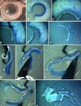
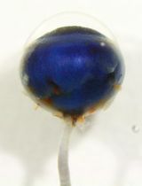
For decades, scientists have tried to identify the mechanisms underlying the differentiation potential of mESCs with general (e.g., Embryoïd Body) or directed (e.g., Neuronal Precursor Cells) differentiation protocols. Recently, the 2i culture media was discovered allowing the captivation of the naïve stem cells state in vitro (Ying et al., 2008). Betschinger and colleagues took advantage of the medium and developed a new ‘exit from pluripotency’ assay allowing the identification of novel factors involved in the commitment of mESCs (Betschinger et al., 2013). Briefly, mESCs maintained in 2i media conditions, are placed for two days in permissive media. Subsequently, the 2i media is reintroduced allowing only the survival of naïve mESCs. In this assay, wild type (WT) mESCs commit to differentiation during the two days of permissive media and die after reintroduction of 2i medium. Indeed, indicating that only two days of permissive media are sufficient to break-down the pluripotency network and commit to differentiation. Unfortunately, mutant mESCs for RNA interference pathways, i.e., Dicer and Dgcr8 genes, proliferate much slower than their WT counterparts making the assessment of the exit from pluripotency in two days with the original protocol less suitable. We decided to extend the presence of the cells in the permissive media to four days and then to reintroduce the 2i media for three more days. Only cells that do not commit during the four days in permissive media conditions will be able to survive and proliferate during the final three days in 2i media. Finally, cell survival and stemness are measured with Alkaline Phosphatase (AP) staining, as in the initial protocol.
Materials and Reagents
- 10 cm plate (TPP Techno Plastic Products, catalog number: 93100 )
- 6-well plate (TPP Techno Plastic Products, catalog number: 92006 )
- 10 ml pipettes (Bioswisstec, catalog number: 515210 )
- 5 ml pipettes (Bioswisstec, catalog number: 515205 )
- 15 ml Falcon tube (Greiner Bio One International, catalog number: 188271 )
- Glass Pasteur pipette (HUBERLAB, catalog number: 1.1127.01 )
- Mouse embryonic stem cells (E14TG2a mESC line obtained from ATCC: ES-E14TG2a) (ATCC, catalog number: CRL-1821 )
- Phosphate-buffered saline (PBS) 1x (Thermo Fisher Scientific, GibcoTM, catalog number: 10010015 )
- Trypsin-EDTA 0.05% (Thermo Fisher Scientific, GibcoTM, catalog number: 25300054 )
- Alkaline phosphatase Kit (Sigma-Aldrich, catalog number: 86R-1KT )
- ddH2O (Sigma-Aldrich, catalog number: 99053 )
Note: This product has been discontinued. - Gelatin (Sigma-Aldrich, catalog number: G1890 )
- Dulbecco’s modified Eagle medium (DMEM) high glucose (Sigma-Aldrich, catalog number: D6429 )
- Fetal bovine serum (FBS) (Thermo Fisher Scientific, GibcoTM, catalog number: 10270106 )
- Leukemia inhibitory factor (LIF) 10 millions unit/ml (Merck, catalog number: ESG1107 )
- 2-Mercaptoethanol (βM) 50 mM (Thermo Fisher Scientific, GibcoTM, catalog number: 31350010 )
- Mixture of penicillin and streptomycin (PS) (Sigma-Aldrich, catalog number: P0781 )
- N2B27 (Takara Bio, catalog number: Y40002 )
- MAPK/ERK inhibitor 0.4 μM (PD03) (Stemolecule PD0325901) (STEMCELL Technologies, catalog number: 72184 )
- GSK3β inhibitor 3 μM (Chiron) (Stemolecule CHIR99021) (STEMCELL Technologies, catalog number: 72054 )
- Acetone (Merck, catalog number: 1.00014.1000 )
- Formaldehyde 37% (Sigma-Aldrich, catalog number: 47608-1L-F )
- 0.2% gelatin solution (see Recipes)
- MES medium (see Recipes)
- MEF medium (see Recipes)
- 2i + LIF medium (see Recipes)
- Fixative solution (see Recipes)
- Substrate solution (see Recipes)
Equipment
- HeracallTM 150i incubator (37 °C and 8% CO2) (Thermo Fischer Scientific, Thermo ScientificTM, model: HeracallTM 150i )
- Centrifuge (Eppendorf, model: 5810 )
- Tissue culture hood (FASTER, model: Safe FAST Premium 209, catalog number: F00024900000 )
- Millipore ScepterTM 2.0 cell counter (EMD Millipore, model: ScepterTM 2.0, catalog number: PHCC00000 ) (or any cell counter)
- Inverted microscope (Nikon Instruments, model: Eclipse TS100 )
Software
- ImageJ software 1.48v
- Adobe Photoshop CS6
- GraphPad Prism 6.0a software
Procedure
文章信息
版权信息
© 2017 The Authors; exclusive licensee Bio-protocol LLC.
如何引用
Readers should cite both the Bio-protocol article and the original research article where this protocol was used:
- Cirera-Salinas, D. and Ciaudo, C. (2017). Exit from Pluripotency Assay of Mouse Embryonic Stem Cells. Bio-protocol 7(16): e2507. DOI: 10.21769/BioProtoc.2507.
- Cirera-Salinas, D., Yu, J., Bodak, M., Ngondo, R. P., Herbert, K. M. and Ciaudo C. (2017). Non-canonical function of DGCR8 controls mESCs exit from pluripotency. J Cell Biol.
分类
干细胞 > 多能干细胞 > 细胞诱导
细胞生物学 > 细胞分离和培养 > 细胞生长
您对这篇实验方法有问题吗?
在此处发布您的问题,我们将邀请本文作者来回答。同时,我们会将您的问题发布到Bio-protocol Exchange,以便寻求社区成员的帮助。