

Image-Based Analysis of Mitochondrial Area and Counting from Adult Mouse Dopaminergic Neurites
基于图像分析成熟小鼠多巴胺能神经元突起中线粒体的面积和数量
发布: 2018年08月20日第8卷第16期 DOI: 10.21769/BioProtoc.2471 浏览次数: 9410
评审: Anonymous reviewer(s)
参见作者原研究论文
The authors used this protocol in:
Jan 2018
Advertisement


Abstract
Mitochondria form dynamic cytoplasmic networks which undergo morphological changes in order to adapt to cellular stresses and signals. These changes can include alterations in size and number within a given cell. Analysis of the whole network can be a useful metric to assess overall mitochondrial health, particularly in neurons, which are highly sensitive to mitochondrial dysfunction. Here we describe a method which combines immunofluorescence and computerized image analysis to measure mitochondrial morphology (quantification of number, density, and area) in dopaminergic neurites of mice expressing mitochondrially-targeted eYFP.
Keywords: Neurite mitochondria (神经元突起线粒体)Background
Mitochondria are double-membraned organelles present in essentially all the cells of every complex organism. Their main function is to supply the majority of cellular energy as ATP, but they also play roles in apoptosis, buffering intracellular Ca2+, reactive oxygen species production, and regulation of membrane potential (Neupert and Herrmann, 2007; Hamanaka and Chandel, 2010; Shutt and McBride, 2013).
These organelles, often depicted as single “bean-like” structures, are in reality components of a dynamic cytoplasmic network. They can undergo major morphological changes regulated by dynamic processes of membrane fusion and fission, a process believed to be involved in the elimination of dysfunctional organelles through a process called mitophagy. The mitochondrial network can also increase as a response to high cellular energy demand (Sheng, 2017; Devine and Kittler, 2018). The morphology of the mitochondrial network can be altered in response to different stressors, and there is a wide range of possible morphologies which are cell-type, or even cell-compartment dependent (Picard et al., 2013). The localization of mitochondria in the dendrites and axons of neurons, in particular, play a crucial role as they provide, in these specific cells, the energy necessary for synaptic transmission (Chang and Reynolds, 2006; Misgeld and Schwarz, 2017).
Mitochondria localization and dynamics in one particular group of cells, the dopaminergic neurons in the substantia nigra, have been extensively studied during the last decades. These neurons’ projections reach the striatum and their loss causes a depletion of striatal dopamine which is the cause of the classical motor symptoms of Parkinson’s disease (PD) (Dauer and Przedborski, 2003; Braak et al., 2004).
The involvement of mitochondrial dysfunctions in these neurons has been investigated since the 80’s (Kopin and Markey, 1988), but the study of mitochondrial dynamics attracted particular interest especially since the discovery that genes mutated in monogenic forms of Parkinson’s disease (in particular Parkin and PINK1) have an essential role in mitochondrial fission/fusion, mitochondrial transport and mitophagy (Koh and Chung, 2010; Narendra and Youle, 2011).
We recently investigated the consequences of the loss of Parkin in a mouse model of PD in which degeneration of dopaminergic neurons was caused by mtDNA depletion and mitochondrial dysfunction (Pinto et al., 2018). In this context, we also found that lack of Parkin affected mitochondrial morphology in dopaminergic axons.
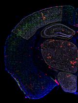
A challenge in studying the mitochondria morphology in vivo is the clear visualization of the organelles and in discerning the ones present in the axons. To study mitochondrial morphology in dopaminergic neurons, we used immunofluorescence microscopy on mice specifically expressing eYFP in the mitochondria of dopaminergic neurons, as well as computerized image analysis software to study mitochondrial number, density and size, as a measure of mitochondrial health (Pinto et al., 2018).
Materials and Reagents
- 500 ml Vacuum Filter (Corning, catalog number: 431097 )
- Single edge razor blades (VWR, catalog number: 55411-050 )
- 24-well Tissue Culture Plate (VWR, catalog number: 10062-896 )
- Microscope Cover Glasses (VWR, catalog number: 16004-098 )
- Micro Slides, Superfrost Plus (VWR, catalog number: 48311-703 )
- Syringes (Insulin Syringes) (BD, catalog number: 328468 )
- Mouse strains: males mito-eYFP DAT-tTA were analyzed at 4 months of age
- Super glue (Henkel, Loctite, catalog number: 1399967 )
- Ketamine (Ketamine HCl, Hospira, catalog number: 02051-05 )
- Xylazine (AnaSed, NDC 59599-110-20)
- Reagent Alcohol (Sigma-Aldrich, catalog number: 793213 )
- Paraformaldehyde (Sigma-Aldrich, catalog number: 441244 )
- 10x PBS (Merck, Calbiochem, catalog number: 6505-4L )
- Hard Set Fluorescent Mounting Medium (Vector Laboratories, catalog number: H-1400 )
- Sodium azide (Sigma-Aldrich, catalog number: S8032 )
- 1x PBS (see Recipes)
- 4% Paraformaldehyde (see Recipes)
- 0.02% (w/v) Sodium azide (see Recipes)
Equipment
- Perfusion machine (Gilson, model: MINIPULS® 3 )
- 23 G Surshield Safety Winged Infusion Set (Terumo Medical, catalog number: SV*S23BL )
- Blunt scissors (Thermo Fisher Scientific, catalog number: 78702 )
- Sharp scissors (Fisher Scientific, catalog number: 13-808-2 )
- Forceps (Fisher Scientific, catalog number: 13-812-39 )
- Metal Spatula (Fisher Scientific, catalog number: 14-374 )
- Acrylic Brain Matrix for Adult Mouse, Coronal Slices, 1 mm spacing (World Precision Instruments, catalog number: RBMA-200C )
- Vibratome (Leica Biosystems, model: Leica VT1000 S )
- Confocal Microscope (ZEISS, Laser Scanning Microscope LSM 710 Observer Z1)
Software
- Zen Software Package (ZEN 2010B SP1, Version 6,0,0,485, Configuration 5.00.01)
- FIJI (FIJI Is Just ImageJ) (Version 2.0.0–rc–67/1.52d)
Notes:- It is a distribution package of ImageJ with plugins for image analysis
- It is available for free download here (https://imagej.net/Fiji/Downloads)
- Microsoft® Excel
Procedure
文章信息
版权信息
© 2018 The Authors; exclusive licensee Bio-protocol LLC.
如何引用
Readers should cite both the Bio-protocol article and the original research article where this protocol was used:
- Nissanka, N., Moraes, C. T. and Pinto, M. (2018). Image-Based Analysis of Mitochondrial Area and Counting from Adult Mouse Dopaminergic Neurites. Bio-protocol 8(16): e2471. DOI: 10.21769/BioProtoc.2471.
- Pinto, M., Nissanka, N. and Moraes, C. T. (2018). Lack of Parkin anticipates the phenotype and affects mitochondrial morphology and mtDNA levels in a mouse model of Parkinson's disease. J Neurosci 38(4): 1042-1053.
分类
神经科学 > 细胞机理 > 线粒体
细胞生物学 > 细胞成像 > 固定组织成像
您对这篇实验方法有问题吗?
在此处发布您的问题,我们将邀请本文作者来回答。同时,我们会将您的问题发布到Bio-protocol Exchange,以便寻求社区成员的帮助。