

Protein Localization in the Cyanobacterium Anabaena sp. PCC7120 Using Immunofluorescence Labeling
免疫荧光标记定位蓝藻鱼腥藻属PCC7120中的蛋白质


(*contributed equally to this work) 发布: 2017年06月05日第7卷第11期 DOI: 10.21769/BioProtoc.2318 浏览次数: 8322
评审: Dennis NürnbergElizabeth LibbyAnonymous reviewer(s)
参见作者原研究论文
The authors used this protocol in:

Mar 2016
Advertisement


Abstract
Techniques such as immunoflorescence are widely used to determine subcellular distribution of proteins. Here we report on a method to immunolocalize proteins in Anabaena sp. PCC7120 with fluorophore-conjugated antibodies by fluorescence microscopy. This method improves the permeabilization of cyanobacterial cells and minimizes the background fluorescence for non-specific attachments. In this protocol, rabbit antibodies were raised against the synthetic peptide of CyDiv protein (Mandakovic et al., 2016). The secondary antibody conjugated to the fluorophore Alexa488 was used due to its different emission range in comparison to the autofluorescence of the cyanobacterium.
Keywords: Cell division (细胞分裂)Background
The immunofluorescence of cyanobacteria has been used extensively in cell identification and counting studies (Jin et al., 2016). However, immunolocalization of proteins has not been achieved efficiently in cyanobacteria. The most recurrent method to localize proteins is by fusing the protein of interest to a fluorescent protein such as GFP (Green Fluorescent Protein) that has a different emission wavelength (compared with cyanobacterial autofluorescence), and subsequent visualization using epifluorescence or confocal microscopy (Flores et al., 2016; Santamaria-Gomez et al., 2016).
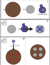
The structural properties of cyanobacterial cells are the main challenges for applying immunofluorescence techniques. They consist of an inner membrane (IM), a peptidoglycan layer (PG) and an outer membrane (OM) (Rippka, 1988; Baulina, 2012; Jin et al., 2016), with an additional exopolysaccharide layer (sheath). The sheath is found in both unicellular and filamentous cyanobacteria (Kehr and Dittmann, 2015), and their thickness, composition and appearance depend on growth conditions, metabolic status, cell differentiation and other external and internal parameters (Jin et al., 2016). The sheath tends to trap antibodies by unspecific interactions. To avoid this problem, the washing and membrane permeabilization steps are the key to a successful immunofluorescence technique in cyanobacteria.
Materials and Reagents
- Pipette tips
- 1.5 ml tubes (Eppendorf)
- 50 ml tubes (Falcon tubes)
- Poly-L-lysine coated glass slides (Sigma-Aldrich, catalog number: P0425-72EA )
- Cover slips
- Petri dish
- Filter with a pore size of 0.2 µm
- Filamentous cyanobacterium, Anabaena sp. PCC7120
- BG-11 liquid supplied with 10 mM NaHCO3 (Rippka, 1988)
- Sodium hydrogen carbonate (NaHCO3) (EMD Millipore, catalog number: 106329 )
- Ethanol (EMD Millipore, catalog number: 1.00983.2500 )
- Triton X-100 (Winkler Limitada, catalog number: BM-2020 )
- Bovine serum albumin (BSA) (Divbio Science, catalog number: 41-903-100 )
- Tween-20 (Winkler Limitada, catalog number: TW-1652 )
- Secondary antibody Alexa Fluor 488 goat anti-rabbit IgG (Thermo Fisher Scientific, Invitrogen, catalog number: A11008 )
- ProLong Gold Antifade Mountant (Thermo Fisher Scientific, InvitrogenTM, catalog number: P36930 )
- Nail varnish
- Primary polyclonal antibody against All2320 peptide (Mandakovic et al., 2016)
- Sodium chloride (NaCl) (EMD Millipore, catalog number: 106404 )
- Potassium chloride (KCl) (EMD Millipore, catalog number: 104938 )
- Sodium dihydrogen phosphate (Na2HPO4) (EMD Millipore, catalog number: 106559 )
- Potassium phosphate monobasic (KH2PO4) (EMD Millipore, catalog number: 529568 )
- PBS buffer (pH 7.4) (see Recipes)
Equipment
- Pipettes
- Hydrophobic PAP pen (Thermo Fisher Scientific, catalog number: 008877 )
- Freezer at -20 °C
- Incubator at 4 °C
- Incubator at 55 °C
- Incubator at 24 °C with white light
- Olympus Fluoview FV1000 Confocal Microscope (Olympus, model: FluoviewTM FV1000 ) and objectives of 60x/1.35 NA oil immersion and 100x/1.40 NA oil immersion. Laserline Argon 488 (Excitation 495 nm, Emission 509 nm) and Laserline DPSS (Excitation 565 nm, Emission 590 nm)
- Moisture chamber (A dark plastic box with a moistened paper inside, PolarSafeTM Polypropylene Freezer Storage Box) (Argos Technologies, catalog number: R3130 )
Software
- ImageJ software (https://imagej.net)
Procedure
文章信息
版权信息
© 2017 The Authors; exclusive licensee Bio-protocol LLC.
如何引用
Trigo, C., Andrade, D. and Vásquez, M. (2017). Protein Localization in the Cyanobacterium Anabaena sp. PCC7120 Using Immunofluorescence Labeling. Bio-protocol 7(11): e2318. DOI: 10.21769/BioProtoc.2318.
分类
微生物学 > 微生物细胞生物学 > 细胞成像
细胞生物学 > 细胞成像 > 荧光
您对这篇实验方法有问题吗?
在此处发布您的问题,我们将邀请本文作者来回答。同时,我们会将您的问题发布到Bio-protocol Exchange,以便寻求社区成员的帮助。