

Single-molecule Analysis of DNA Replication Dynamics in Budding Yeast and Human Cells by DNA Combing
采用DNA梳理技术对芽殖酵母和人类细胞中DNA复制动力学进行单分子分析

发布: 2017年06月05日第7卷第11期 DOI: 10.21769/BioProtoc.2305 浏览次数: 15064
评审: Emilie BesnardAnonymous reviewer(s)
参见作者原研究论文
The authors used this protocol in:
Jun 2012
Advertisement


Abstract
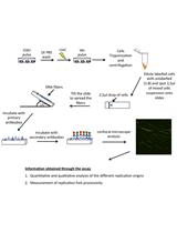
The DNA combing method allows the analysis of DNA replication at the level of individual DNA molecules stretched along silane-coated glass coverslips. Before DNA extraction, ongoing DNA synthesis is labeled with halogenated analogues of thymidine. Replication tracks are visualized by immunofluorescence using specific antibodies. Unlike biochemical and NGS-based methods, DNA combing provides unique information on cell-to-cell variations in DNA replication profiles, including initiation and elongation. Finally, this assay can be used to monitor the effect of DNA lesions on fork progression, arrest and restart.
Keywords: Replication (复制)Background
DNA synthesis is initiated at thousands of sites on eukaryotic chromosomes called replication origins. Origin activation follows a well-defined replication timing program that is controlled by checkpoint kinases and epigenetic modifications of chromatin (Prioleau and MacAlpine, 2016). Replication forks frequently stall during a normal S phase. Fork arrest is caused by multiple events, such as DNA lesions, tightly bound protein complexes, and transcription at highly expressed genes (Tourriere and Pasero 2007; Zeman and Cimprich, 2013). Eukaryotes have developed different strategies to deal with this replication stress, including repair mechanisms to restart arrested forks and activation of dormant replication origins to rescue terminally-arrested forks.
DNA combing is a method of choice to monitor different aspects of replication (fork speed, origin usage, fork restart, sister fork asymmetry). Unlike other DNA fiber methods such as DNA fiber spreading, the stretching, density and alignment of DNA molecules are highly reproducible and tightly controlled in the DNA combing method. Stretching is imposed by the force exerted by a receding air/water interface, independently of the length of DNA fibers (Bensimon et al., 1994; Michalet et al., 1997). Origin firing and progression of replication forks are followed after incorporation of thymidine analogs, such as 5-bromo-2’-deoxyuridine (BrdU), 5-iodo-2’-deoxyuridine (IdU) and 5-chloro-2’-deoxyuridine (CldU) in newly-synthesized DNA. This technique has been successfully used to monitor DNA replication dynamics in a variety of organisms, including bacteria, yeast, Drosophila, Xenopus and mammals.
Here, we provide detailed protocols to analyze newly synthesized DNA fibers in budding yeast and in human cells and to investigate various aspects of DNA replication in normal growth conditions and under replicative stress.
Materials and Reagents
- Common to human/yeast cells
- Tape
- 14 ml round-bottom polypropylene tubes (Corning, Falcon®, catalog number: 352059 )
- Tips 1 ml, 200 µl, 20 µl
- Silanized coverslips (Genomic Vision, catalog number: COV-001 ) purchased from Genomic Vision or prepared as described (Labit et al., 2008)
- 2 ml Teflon reservoir (Reservoir MCS Support [x 2]; from Genomic Vision)
- Whatman paper
- Microscope slides SuperFrost (VWR, catalog number: 630-1987 )
- Cyanoacrylate glue
- Diamond tip engraving pen (Sigma-Aldrich, catalog number: Z225568-1EA )
- Saran plastic film (Dominique Dutscher, catalog number: 090264 )
- Coplin Jar
- EDTA (Sigma-Aldrich, catalog number: E6758 )
- LMP agarose (Bio-Rad Laboratories, catalog number: 161-3111 )
- Plug mold (Bio-Rad Laboratories, catalog number: 170-3713 )
- Proteinase K (Sigma-Aldrich, catalog number: P6556 )
- 10x PBS (Sigma-Aldrich, catalog number: D1408 )
- YOYO-1 (Thermo Fisher Scientific, InvitrogenTM, catalog number: Y3601 )
- Sodium chloride (NaCl) (VWR, catalog number: 27810-295 )
- β-agarase (New England Biolabs, catalog number: M0392L )
- Sodium hydroxide (NaOH) (Merck, catalog number: 1.06462.1000 )
- BSA fraction V (Sigma-Aldrich, catalog number: A9647 )
- Prolong Gold Antifade reagent (Thermo Fisher Scientific, InvitrogenTM, catalog number: P36930 )
- BrdU (Sigma-Aldrich, catalog number: B5002 )
- IdU (Sigma-Aldrich, catalog number: I7125 )
- CldU (MP Biomedicals, catalog number: 0 2105478 )
- DMSO (Sigma-Aldrich, catalog number: D2650 )
- Hydroxyurea (Sigma-Aldrich, catalog number: H8627 )
- N-laurylsarcosine sodium salt (Sigma-Aldrich, catalog number: L9150 )
- MES hydrate (Sigma-Aldrich, catalog number: M2933 )
- MES sodium salt (Sigma-Aldrich, catalog number: M5057 )
- Triton X-100 (Sigma-Aldrich, catalog number: T8787 )
- Mouse anti-BrdU clone B44 IgG1 (BD, BD Biosciences, catalog number: 347580 )
- Rat anti-BrdU clone BU1/75 (Bio-Rad Laboratories, catalog number: OBT0030 )
- Mouse anti ssDNA (poly dT) IgG2a (EMD Millipore, catalog number: MAB3034 or MAB3868 )
- Goat anti-Rat Alexa 488 (Thermo Fisher Scientific, Invitrogen, catalog number: A-11006 )
- Goat anti-Mouse Alexa 546 (Thermo Fisher Scientific, Invitrogen, catalog number: A-11030 )
- Goat anti-Mouse IgG2a Alexa 647 (Thermo Fisher Scientific, Invitrogen, catalog number: A-21241 )
- Goat anti-Mouse IgG1 Alexa 546 (Thermo Fisher Scientific, Invitrogen, catalog number: A-21123 )
- LMP agarose (see Recipes)
- TE50 (see Recipes)
- TE 1x (see Recipes)
- 10x MES buffer pH 6 (see Recipes)
- 1 N NaOH (see Recipes)
- PBS/T (see Recipes)
- Antibodies (dilution in PBS/T) (see Recipes)
- Detection of CldU/IdU/ssDNA (see Recipes)
- Detection of BrdU/ssDNA (see Recipes)
- Tape
- S. cerevisiae specific reagents
- Yeast strain PP872 (genetic background: W303; genotype: MATa, ade2-1, trp1-1, can1-100, leu2-3,112, his3-11,15, ura3, GAL, psi+, RAD5, ura3::URA3-GPD-TK7) (Crabbe et al., 2010)
- Yeast strain HSV-TK + hENT1 (Viggiani and Aparicio, 2006)
- Bacto peptone (BD, BactoTM, catalog number: 211677 )
- Adenine (Sigma-Aldrich, catalog number: A8626 )
- Yeast extract (Sigma-Aldrich, catalog number: 70161 )
- Glucose (VWR, catalog number: 101175P )
- Alpha factor (custom peptide synthesis, Sequence: WHWLQLKPGQPMY)
- Pronase (EMD Millipore, catalog number: 53702-50KU )
- Sodium azide (NaN3) (Sigma-Aldrich, catalog number: 71289 )
- Tris-HCl (Sigma Aldrich, catalog number: RES3098T )
- Citric acid monohydrate (C6H8O7·H2O) (Sigma-Aldrich, catalog number: C1909 )
- Sodium phosphate dibasic (Na2HPO4) (Sigma-Aldrich, catalog number: S3264 )
- Monopotassium phosphate (KH2PO4) (VWR, catalog number: 26936.293 )
- Dibasic potassium phosphate (K2HPO4) (VWR, catalog number: 26930.293 )
- Enzyme powder Zymolyase 20T (MP Biomedicals, catalog number: 0 8320921 )
- YPAD (see recipes)
- Zymolyase buffer (see Recipes)
- 1 M potassium phosphate buffer pH 7.0 (see Recipes)
- 0.1 M citrate phosphate buffer pH 5.6 (see Recipes)
- BrdU stock solution (see Recipes)
- Proteinase K buffer (see Recipes)
- Yeast strain PP872 (genetic background: W303; genotype: MATa, ade2-1, trp1-1, can1-100, leu2-3,112, his3-11,15, ura3, GAL, psi+, RAD5, ura3::URA3-GPD-TK7) (Crabbe et al., 2010)
- Human cells specific reagents
Equipment
- Centrifuge (Eppendorf, model: 5810 R )
- Microcentrifuge (Eppendorf, model: MiniSpin® )
- Thermoblock (thermomixer comfort Eppendorf with 2 ml; 15 ml and 50 ml block)
- Cell counter (CASY® Modell TT - Cell Counter, OLS for yeast cells or Malassez hemocytometer [VWR, catalog number: 631-0975 ] for human cells)
- Pasteur pipette Rubber bulb (Dominique Dutscher, catalog number: 042250 )
- Microwave
- Roller mixer (Cole-Parmer, Stuart, model: SRT9D )
- Leica DM6000B microscope (Leica Microsystems, model: DM6000B )
- CoolSNAP HQ CCD camera (Photometrics, model: CoolSNAP HQ CCD )
- P1000, P200, P20 pipetman
- DNA combing device (Genomic Vision, catalog number: MCS-001 ). Can also be assembled as described (Gallo et al., 2016; Kaykov et al., 2016; Norio and Schildkraut, 2001)
- Metal coverslip holder
- Humid chamber (StainTray slide staining system) (Sigma-Aldrich, catalog number: Z670146-1EA )
- Hybridization oven (Shake ‘n’ StackTM Hybridization Ovens) (Thermo Fisher Scientific, Thermo ScientificTM, catalog number: 6240TS )
- Epifluorescence microscope Zeiss Axio Imager Z2 (Zeiss, model: Axio Imager Z2 ) with a camera Hamamatsu ORCA-Flash4.0 LT, Cmos sensor of 6.5 µm with an objective Zeiss 40x PL APO 1.4 oil and with the filters GFP HE: BP470/40 FT495 BP525/50; Texas Red: BP560/40 FT585 BP630/75; CY5: BP 640/30 FT660 BP690/50)
- Microscope (Nikon Instruments, model: YS100 )
Software
- MetaMorph (Molecular Devices) or open source software such as ImageJ (http://rsbweb.nih.gov/ij/) with bio-formats plugins
- IDeFIx Montpellier (IGMM, CNRS) or software developed by Genomic Vision
- Prism 7.0 (GraphPad) or open source software such as R (https://www.r-project.org/)
Procedure
文章信息
版权信息
© 2017 The Authors; exclusive licensee Bio-protocol LLC.
如何引用
Tourrière, H., Saksouk, J., Lengronne, A. and Pasero, P. (2017). Single-molecule Analysis of DNA Replication Dynamics in Budding Yeast and Human Cells by DNA Combing. Bio-protocol 7(11): e2305. DOI: 10.21769/BioProtoc.2305.
分类
癌症生物学 > 通用技术 > 生物化学试验 > DNA 复制
生物化学 > DNA > 单分子活性 > 成像
分子生物学 > DNA > DNA 标记
您对这篇实验方法有问题吗?
在此处发布您的问题,我们将邀请本文作者来回答。同时,我们会将您的问题发布到Bio-protocol Exchange,以便寻求社区成员的帮助。