

Lung Section Staining and Microscopy
肺切片染色和显微镜观察
发布: 2017年05月20日第7卷第10期 DOI: 10.21769/BioProtoc.2286 浏览次数: 27487
评审: Ivan ZanoniAnonymous reviewer(s)
参见作者原研究论文
The authors used this protocol in:

May 2016
Advertisement


Abstract
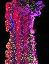
Our protocol describes immunofluorescent staining, hematoxylin and eosin staining and Masson’s trichrome staining on lung sections.
Keywords: Immunofluorescent staining (免疫荧光染色)Background
The primary function of the lungs is gas exchange. The lungs are composed of various specialized cells and tissues, including the bronchi, the bronchioles and the pulmonary alveoli to facilitate gas exchange. To study lung development or lung diseases in mouse models, the lungs can be removed from mice and either frozen and embedded in optimal cutting temperature (OCT) compound or chemically preserved and embedded in paraffin. To preserve lung tissue architecture, we filled the lung with 1 ml of OCT compound for preparing frozen sections, or 10% buffered formalin for paraffin sections (Zhou et al., 2016). Lung sections are then sliced from frozen or paraffin-embedded lungs and mounted onto slides for preparation of staining. We used frozen tissue sections for antibody-based immunofluorescent staining and used paraffin-embedded sections for hematoxylin and eosin (H&E) staining or Masson’s trichrome staining. Frozen sections are quicker to prepare for immunofluorescent staining and most antibodies work well on frozen sections. Paraffin sections can also be used for immunofluorescent staining, but they require deparaffinization, rehydration and antigen retrieval. Some antibodies do not work well on paraffin sections even after antigen retrieval. However, paraffin samples can be stored at room temperate for very long periods and can be easily cut into very thin sections. Paraffin sections preserve better tissue morphology than frozen sections, so they are better for H&E or trichrome staining.
Immunofluorescent staining is a type of immunohistochemistry that makes use of fluorophores to visualize the location of the antibodies that specifically bind to their target proteins. H&E staining is the most widely used stain in histology and medical diagnosis. This staining method involves application of hemalum and eosin Y that color cell nuclei blue and cytoplasm pink to red. Masson’s trichrome staining is a three-color staining that is used for detecting collagen fibers in tissues. The staining produces blue collagen, dark brown to black cell nuclei and red background.
Materials and Reagents
- Fisherbrand Superfrost Plus microscope slides (Fisher Scientific, catalog number: 12-550-15 )
- Disposable mold
- Coverslip slides
- C57BL/6 mice
- Tissue-Tek OCT compound (SAKURA FINETEK, catalog number: 4583 )
- Formaldehyde, 37-40% ACS (Newcomer Supply, catalog number: 1089 )
- Triton X-100 (Sigma-Aldrich, catalog number: X100 )
- Phosphate buffered saline (PBS, pH 7.4) (Thermo Fisher Scientific, GibcoTM, catalog number: 10010023 )
- Serum from secondary antibody’s host
- Primary antibodies: Rabbit anti α-smooth muscle actin (α-SMA) antibody (Abcam, catalog number: ab5694 )
- Secondary reagents: Texas Red conjugated goat anti-rabbit IgG antibody (Abcam, catalog number: ab6719 )
- Prolong® Gold anti-fade reagent (Cell Signaling Technology, catalog number: 9071 ), or with DAPI (Cell Signaling Technology, catalog number: 8961 )
- Paraffin
- Ethyl alcohol (Newcomer Supply, catalog number: 10841 )
- Xylene (Newcomer Supply, catalog number: 1446 )
- S-mounting medium (Newcomer Supply, catalog number: 6750 )
- Bouin Fluid (Newcomer Supply, catalog number: 1020 )
- Biebrich scarlet-acid fuchsin solution (Sigma-Aldrich, catalog number: HT151 )
- Bovine serum albumin (BSA) (Sigma-Aldrich, catalog number: A3912 )
- Fetal bovine serum (FBS) (GE Healthcare, HycloneTM, catalog number: SH30396.03 )
- Eosin Y (Sigma-Aldrich, catalog number: E4009 )
- Acetic acid, glacial (Sigma-Aldrich, catalog number: A6283 )
- Aluminum potassium sulfate (Sigma-Aldrich, catalog number: 237086 )
- Hematoxylin (Sigma-Aldrich, catalog number: H3136 )
- Sodium iodate (Sigma-Aldrich, catalog number: S4007 )
- Citric acid (Sigma-Aldrich, catalog number: 251275 )
- Ferric chloride (Sigma-Aldrich, catalog number: 157740 )
- Hydrochloric acid (Sigma-Aldrich, catalog number: 320331 )
- Phosphomolybdic acid solution (Sigma-Aldrich, catalog number: HT153 )
- Phosphotungstic acid solution (Sigma-Aldrich, catalog number: HT152 )
- Aniline blue (Sigma-Aldrich, catalog number: 415049 )
- Sodium chloride (NaCl) (Sigma-Aldrich, catalog number: S7653 )
- Sodium phosphate monobasic (NaH2PO4) (Sigma-Aldrich, catalog number: S8282 )
- Blocking buffer (see Recipes)
- Eosin Y solution (see Recipes)
- Eosin Y working solution (0.25%) (see Recipes)
- Mayer’s hematoxylin solution (see Recipes)
- Weigert’s iron hematoxylin solution (see Recipes)
- Phosphomolybdic-phosphotungstic acid solution (see Recipes)
- Aniline blue solution (see Recipes)
Equipment
- Cryostat microtome
- -70 °C freezer
- Fluorescence microscope or a confocal microscope
- Staining jar and slide rack, for example EasyDip Slide Staining System (Newcomer Supply, catalog number: 5300KIT )
- Optical microscope
Procedure
文章信息
版权信息
© 2017 The Authors; exclusive licensee Bio-protocol LLC.
如何引用
Zhou, X. and Moore, B. B. (2017). Lung Section Staining and Microscopy. Bio-protocol 7(10): e2286. DOI: 10.21769/BioProtoc.2286.
分类
免疫学 > 免疫细胞染色 > 免疫检测
细胞生物学 > 细胞成像 > 共聚焦显微镜
您对这篇实验方法有问题吗?
在此处发布您的问题,我们将邀请本文作者来回答。同时,我们会将您的问题发布到Bio-protocol Exchange,以便寻求社区成员的帮助。