

Immunostaining of Formaldehyde-fixed Metaphase Chromosome from Untreated and Aphidicolin-treated DT40 Cells
对照和蚜肠酶素处理的DT40细胞中经甲醛固定的中期染色体的免疫染色

发布: 2017年05月05日第7卷第9期 DOI: 10.21769/BioProtoc.2259 浏览次数: 10702
评审: Antoine de MorreeTatsuki KunohXiaoyi Zheng
参见作者原研究论文
The authors used this protocol in:

Aug 2015
Advertisement


Abstract
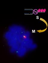
During mitosis chromosomes are condensed into dense X-shaped structures that allow for microscopic determination of karyotype as well as inspection of chromosome morphology.
This protocol describes a method to perform immunostaining of formaldehyde-fixed metaphase chromosomes from the avian cell line DT40. It was developed to characterize the localization of YFP-tagged TopBP1 on mitotic chromosomes and specifically determine the percentage of TopBP1 foci that formed on breaks/gaps as well as ends of individual metaphase macrochromosomes (Pedersen et al., 2015). For this purpose immunostaining of YFP was applied. However, the protocol may be optimized for other cell lines or epitopes.
Background
Microscopic analysis of stained metaphase chromosomes is a classical cytogenetic technique that is extensively used for both research and diagnostics. The basic principle involves induction of cell cycle arrest in metaphase by a spindle destabilizing reagent such as colcemid, which will trigger the spindle assembly checkpoint and therefore arrests cells in metaphase. This serves to enrich for cells with condensed chromosomes. Subsequently cells are subjected to swelling in hypotonic solution followed by spreading of mitotic cells on a microscope slide. The final result is microscopically detectable chromosomes from single cells convenient for karyotype analysis as well as investigations of individual chromosomes. Traditionally, swollen cells are fixed with methanol and acetic acid (3:1) before spreading on slides (Hungerford, 1965; Ronne et al., 1979).
The method described here uses formaldehyde rather than methanol for fixation. This can be useful for subsequent staining with antibodies that are not compatible with methanol fixation. The protocol is optimized for metaphase spreads from chicken DT40 cells, and immunostaining of YFP-tagged TopBP1 on metaphase macrochromosomes (Pedersen et al., 2015). TopBP1 foci on mitotic chromosomes mark DNA insults that are transmitted to G1 daughter cells (Pedersen et al., 2015; Gallina et al., 2016; Oestergaard and Lisby, 2016). The fluorescent signal of YFP is lost during the preparation of metaphase spreads, therefore this protocol includes immunostaining of the YFP epitope. However, it should be possible to apply the protocol for other cell lines and epitopes by optimizing incubation time in hypotonic buffer and antibody concentrations, respectively.
Aphidicolin is a replication inhibitor, which at low concentration induces formation of gaps and breaks on metaphase chromosomes preferentially at common fragile sites (Durkin and Glover, 2007). As stated by this protocol, DT40 cells may be subjected to 0.5 μM aphidicolin to induce breaks and gaps on metaphase chromosomes in DT40 (Pedersen et al., 2015).
The avian karyotype comprises macrochromosomes as well as mini and microchromosomes. The latter two groups are too small to reliably determine features such as breaks/gaps or ends. They are therefore not included in this analysis.
Materials and Reagents
- 15 ml tubes (such as Sigma-Aldrich, catalog number: T1943 )
- Pipette tips
- Round cover slips (Ø12 mm) (Thermo Fisher Scientific)
- Cytospin slides (Thermo Fisher Scientific, catalog number: 5991059 )
- A DT40 cell line (Baba and Humphries, 1985) carrying endogenous YFP-tagging at the TopBP1 gene (Germann et al., 2014; Pedersen et al., 2015)
- Dimethyl sulfoxide (DMSO) (Sigma-Aldrich, catalog number: D4540 )
- Aphidicolin (Sigma-Aldrich, catalog number: A0781 )
- Colcemid (Thermo Fisher Scientific, GibcoTM, catalog number: 15212012 )
- Cytofunnels (Thermo Fisher Scientific, Thermo ScientificTM, catalog number: A78710020 )
- Nail polish
- RPMI 1640 medium GlutaMAX (Thermo Fisher Scientific, GibcoTM, catalog number: 61870044 )
- Chicken serum (such as Sigma-Aldrich, catalog number: C5405 or Thermo Fisher Scientific, Thermo ScientificTM, catalog number: 16110082 )
- Fetal bovine serum (FBS) (Heat inactivated) (Thermo Fisher Scientific, GibcoTM, catalog number: 10500 )
- β-mercaptoethanol (Sigma-Aldrich, catalog number: M6250 )
- Penicillin/streptomycin (Thermo Fisher Scientific, GibcoTM, catalog number: 15140122 )
- Potassium chloride (KCl) (TH GEYER, Chemsolute®, catalog number: 1632 )
- Paraformaldehyde (Sigma-Aldrich, catalog number: 158127 )
- Sodium hydroxide (NaOH)
- Hydrochloric acid (HCl)
- Sodium chloride (NaCl) (Avantor® Performance Materials, J.T. Baker, catalog number: 0278.1000 )
- 2-Amino-2-(hydroxymethyl)-1,3-propanediol (Tris, Trizma base) (Sigma-Aldrich, catalog number: T1503 )
- (Ethylenedinitrilo)tetraacetic acid (EDTA) (Sigma-Aldrich, catalog number: 27285 )
- Triton X-100 (Sigma-Aldrich, catalog number: X100 )
- Tween-20 (Sigma-Aldrich, catalog number: P9416 )
- Bovine serum albumin (BSA) (Sigma-Aldrich, catalog number: A4503 )
- Alexa Fluor 488-conjugated anti-mouse IgG (Thermo Fisher Scientific, Invitrogen, catalog number: A21121 )
- Mouse anti-GFP antibody (Roche Diagnostics, catalog number: 11814460001 )
- Glycerol (AppliChem, catalog number: 142329.1211 )
- n-propyl-gallate (Sigma-Aldrich, catalog number: 02370 )
- 2-(4-Amidinophenyl)-6-indolecarbamidine dihydrochloride, 4’,6-Diamidino-2-phenylindole dihydrochloride (DAPI) (Sigma-Aldrich, catalog number: D9542 )
- DT40 cells medium (see Recipes)
- Aphidicolin solution (see Recipes)
- Hypotonic buffer (see Recipes)
- 3% paraformaldehyde in PBS (see Recipes)
- KCM buffer (see Recipes)
- Phosphate buffered saline (PBS) (see Recipes)
- PBS-T (see Recipes)
- Blocking solution (see Recipes)
- Alexa 488-secondary antibody working solution (see Recipes)
- Anti-GFP antibody working solution (see Recipes)
- DAPI-mounting buffer (see Recipes)
Equipment
- Cytocentrifuge, Cytospin4 (Thermo Fisher Scientific, Thermo ScientificTM, model: CytospinTM 4 Cytocentrifuge )
- Pipettes (P1000, P200, P20)
- CO2 incubator (such as Nuaire, model: 4750E , series 6)
- Flasks such as 25 cm2 flasks (TPP, catalog number: 90025 )
- Vortex
- Fume hood
- Coplin jar such as ‘jar staining acc to Coplin W/cover’ (VWR, catalog number: 631-9331 )
- Fluorescence microscope (such as GE Healthcare, DeltaVision Elite; Applied Precision) equipped with a 100x objective lens (NA 1.4; Olympus, model: U-PLAN S-APO ), a cooled EM CCD camera (Photometrics, model: Evolve 512 ), and a solid-state illumination source (Insight; Applied Precision) or similar fluorescence microscope. The microscope should be equipped with appropriate filters to image Alexa488 and DAPI
Software
- SoftWoRx (Applied Precision) software or similar
- Volocity software (PerkinElmer) or ImageJ
Procedure
文章信息
版权信息
© 2017 The Authors; exclusive licensee Bio-protocol LLC.
如何引用
Readers should cite both the Bio-protocol article and the original research article where this protocol was used:
- Oestergaard, V. H. (2017). Immunostaining of Formaldehyde-fixed Metaphase Chromosome from Untreated and Aphidicolin-treated DT40 Cells. Bio-protocol 7(9): e2259. DOI: 10.21769/BioProtoc.2259.
-
Pedersen, R. T., Kruse, T., Nilsson, J., Oestergaard, V. H. and Lisby, M. (2015). TopBP1 is required at mitosis to reduce transmission of DNA damage to G1 daughter cells. J Cell Biol 210(4): 565-582.
分类
癌症生物学 > 通用技术 > 细胞生物学试验 > 细胞周期
细胞生物学 > 细胞染色 > 核酸
您对这篇实验方法有问题吗?
在此处发布您的问题,我们将邀请本文作者来回答。同时,我们会将您的问题发布到Bio-protocol Exchange,以便寻求社区成员的帮助。