

Automated Tracking of Root for Confocal Time-lapse Imaging of Cellular Processes
对根进行自动跟踪以进行细胞过程的共焦延时成像


(*contributed equally to this work) 发布: 2017年04月20日第7卷第8期 DOI: 10.21769/BioProtoc.2245 浏览次数: 11796
评审: Tie LiuShahin S. AliIsabelle Colas
参见作者原研究论文
The authors used this protocol in:

Jun 2016
Advertisement


Abstract
Here we describe a protocol that enables to automatically perform time-lapse imaging of growing root tips for several hours. Plants roots expressing fluorescent proteins or stained with dyes are imaged while they grow using automatic movement of the microscope stage that compensates for root growth and allows to follow a given region of the root over time. The protocol makes possible the image acquisition of multiple growing root tips, therefore increasing the number of recorded mitotic events in a given experiment. The protocol also allows the visualization of more than one fluorescent protein or dye simultaneously, using multiple channel acquisition. We particularly focus on imaging of cytokinesis in Arabidopsis root tip meristem, but this protocol is also suitable to follow root hair growth, pollen tube growth, and other regions of root over time, in various plant species. It may as well be amenable to automatically track non-plant structures with an apical growth.
Keywords: Cell division (细胞分裂)Background
Cytokinesis is the last step of cell division, when the mother cell cytoplasm is partitioned between two daughter cells (Lipka et al., 2015). In plants, it is achieved through the centrifugal expansion of a cell plate in the division plane, which eventually becomes the newly synthetized cell wall between the cells that underwent mitosis (Buschmann and Zachgo, 2016; Müller and Jürgens, 2016). Plant cells, being embedded in a stiff cell wall, cannot migrate. Orientation of cell division together with elongation is therefore critical for organ morphogenesis. Root meristems are a good model to study cell division because they are easily amenable to microscopy techniques without the need of dissection. However, roots undergoing cell division grow in length, and therefore require manual adjustment of the observation field over time. This protocol allows easy time-lapse imaging of cytokinesis, and of other cellular processes.
Materials and Reagents
- 12-well microplates (Corning, Costar®, catalog number: 3513 )
- Microscope slides 76 x 26 x 1.1 mm (RS Components, catalog number: ISO 8037 )
- Microscope coverslips 22 x 60 mm (Thermo Fisher Scientific, Menzel-Gläser, catalog number: 630-2102 )
- Observation chambers, Lab-Tek II Chambered Coverglass W/Cover #1.5 Borosilicate (Thermo Fisher Scientific, Thermo ScientificTM, catalog number: 155360 )
- Strait scalpel blades (e.g., Swann Morton, straights mounted BS EN 27740 blades)
- Arabidopsis thaliana, 4 to 7 days-old seedlings of wild-type genotype, or expressing membrane and/or cell plate-localized fluorescent fusion proteins (e.g., 2xp35S::MP:YFP in Col-0, where MP is a myristoylation and palmitoylation signal sequence [Martinière et al., 2012; Simon et al., 2016]). Seedling can be grown vertically in squared plates (90 x 90 x 15) or round plates to get intact growing roots
- Murashige and Skoog (MS) medium (Sigma-Aldrich, catalog number: M5519 )
- Agar (Sigma-Aldrich, catalog number: A7921-1KG )
- FM4-64 (N-(3-Triethylammoniumpropyl)-4-(6-(4-(Diethylamino) Phenyl) Hexatrienyl) Pyridinium Dibromide) (Thermo Fisher Scientific, Molecular ProbesTM, catalog number: T-3166 )
- Suitable fluorescent dyes (alternative) or fluorescent proteins for registration
- Plasma membrane or cell wall dyes such as FM4-64 or propidium iodide (Sigma-Aldrich, catalog number: P4864 )
- Nuclei dyes such as Hoechst (Thermo Fisher Scientific, InvitrogenTM, catalog number: H21486 )
- Plasma membrane or cell wall dyes such as FM4-64 or propidium iodide (Sigma-Aldrich, catalog number: P4864 )
Equipment
- Inverted confocal microscope (e.g., Carl Zeiss, model: AxioObserver Z1 ), equipped with a spinning disk module (e.g., Yokogawa Electric, model: CSU-W1 [T3 model])
- Metamorph and ImageJ software on the computer connected to the microscope
- 20 °C plant growth chamber with 24 h daylight (e.g., SANYO Electric, model: MLR-351 )
- Small tweezers (e.g., straight fine tweezers)
- Sterile hood
Software
- ImageJ (https://imagej.nih.gov/ij/; Schneider et al., 2012; this protocol was tested with version 1.49p but should be compatible with newer versions) with the plugins Turboreg (P. Thévenaz, http://bigwww.epfl.ch/thevenaz/turboreg/) and Stackreg (P. Thévenaz, http://bigwww.epfl.ch/thevenaz/stackreg/) installed are required. Fiji (http://fiji.sc/; Schindelin et al., 2012) could be used instead of ImageJ because it bundles the required plugins. To add the plugins, simply download them and copy them in ImageJ/plugins folder.
- Download the three files (CL_ini-Pos-Ch-finished.JNL; CL_ini-Pos-Ch.JNL; CL-root-track-MM_63multiD.ijm) attached as a zip file (named Files_BioProtocol_Doumane, as well available at this url http://www.ens-lyon.fr/RDP/SiCE/METHODS.html) and extract them in the folder C:\MM\app\mmproc\journal\root-tracking\. The ImageJ macro file CL-root-track-MM_63multiD.ijm should be edited according to the calibration of the objective used. Open the file in ImageJ (use the menu Open>File...), search for the line with ‘objective’, and replace the number 0.2063 with the real size of the pixel in microns, on this line and the next one (when using our 63x objective, 1 pixel is 0.2063 microns wide; Figure 1; Note 5). Save the file. Install the macro by selecting it in Plugins > Macro > Install. CL-root-track-MM_63multiD should appear in the Plugins > Macros > lower panel (using a 63x objective).
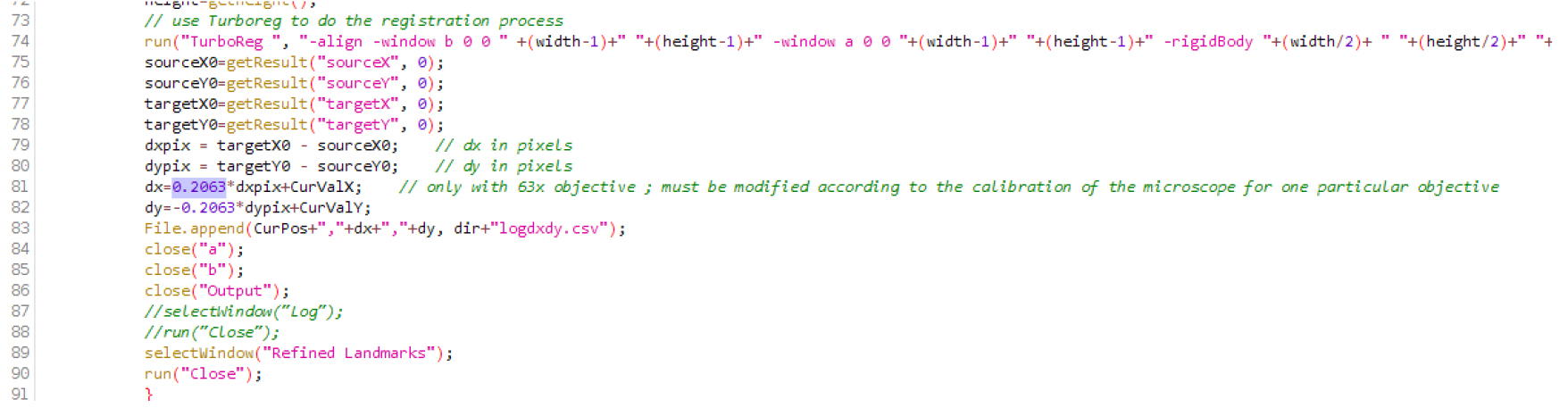
Figure 1. Macro file .ijm in the Fiji editor. Replace the number 0.2063 (here in lines 81 and 82) by the real size of the pixel (in microns) on your microscope, with the objective that will be used.
Procedure
文章信息
版权信息
© 2017 The Authors; exclusive licensee Bio-protocol LLC.
如何引用
Doumane, M., Lionnet, C., Bayle, V., Jaillais, Y. and Caillaud, M. (2017). Automated Tracking of Root for Confocal Time-lapse Imaging of Cellular Processes. Bio-protocol 7(8): e2245. DOI: 10.21769/BioProtoc.2245.
分类
植物科学 > 植物发育生物学 > 形态建成
细胞生物学 > 细胞成像 > 共聚焦显微镜
您对这篇实验方法有问题吗?
在此处发布您的问题,我们将邀请本文作者来回答。同时,我们会将您的问题发布到Bio-protocol Exchange,以便寻求社区成员的帮助。