

Physical Removal of the Midbody Remnant from Polarised Epithelial Cells Using Take-Up by Suction Pressure (TUSP)
采用吸压抽吸法(TUSP)物理去除极化上皮细胞中的中间体残留物

发布: 2017年04月20日第7卷第8期 DOI: 10.21769/BioProtoc.2244 浏览次数: 8992
评审: Andrea PuharJingli CaoThirupugal Govindarajan
参见作者原研究论文
The authors used this protocol in:
Aug 2016
Advertisement


Abstract
In polarised epithelial cells the midbody forms at the apical cell surface during cytokinesis. Once severed, the midbody is inherited by one of the daughter cells remaining tethered to the apical plasma membrane where it participates in non-cytokinetic processes, such as primary ciliogenesis. Here, we describe a novel method to physically remove the midbody remnant from cells and assess the possible effects caused by its loss (Bernabé-Rubio et al., 2016).
Keywords: Epithelial cells (上皮细胞)Background
The midbody or the Flemming body is the central part of the intercellular bridge formed between daughter cells during the final stages of mitosis. The abscission on either side of the bridge by the endosomal sorting complexes required for transport (ESCRT) machinery, results in the physical separation of the two daughter cells (Green et al., 2012). In addition to its known function in the regulation of mitosis, recent studies have begun to elucidate post-mitotic roles for the midbody. Due to its role in the initiation of lumen formation in kidney cells, the midbody has been postulated to serve as a polarity cue (Li et al., 2014). More recently, it has been demonstrated that the midbody remnant is directly involved in primary ciliogenesis by polarised Madin-Darby canine kidney (MDCK) cells (Bernabé-Rubio et al., 2016). It has been also found to have a role in formation of the dorsoventral axis during the development of Caenorhabditis elegans (Singh and Pohl, 2014), and in defining cell fate and differentiation (Kuo et al., 2011). Previous studies have used laser ablation to impair the function of the midbody remnant. When performed in cultured cell lines, however, laser ablation can result in cell death due to damage of the plasma membrane and proximal cytosolic elements. Accordingly, we have designed a gentle procedure, which we have called ‘take-up by suction pressure’ (TUSP). TUSP allows non-deleterious midbody remnant removal from the cell surface of epithelial cells. The fundamental principle is based on using a fine-aperture glass pipette attached to patch-clamp apparatus to physically remove the midbody with applied negative pressure (Figure 1).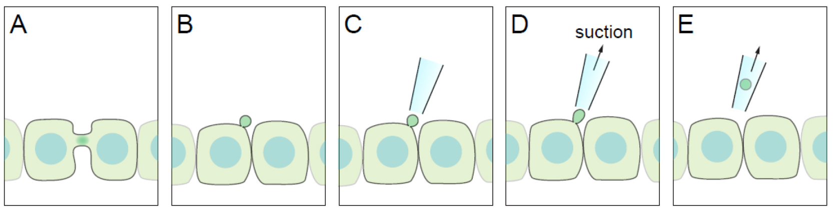
Figure 1. Diagram of the TUSP procedure. A. An apical intercellular bridge forms during cytokinesis in polarised epithelial cells. B. After abscission, one of the daughter cells inherits the midbody as a remnant, which will be positioned over the apical cell surface. C-E. By using a glass pipette connected to path-clamp apparatus, the midbody remnant can be removed from cells if suction pressure is applied.
Materials and Reagents
- 12 mm glass coverslips #1 (VWR, catalog number: 631-0713 )
- Gridded coverslips (optional) (Electron Microscopy Sciences, catalog number: 72265-12 )
- Falcon 24-well plates (Corning, catalog number: 353047 )
- Permanent marker (Faber-Castell Multimark 1523) (CultPens, catalog number: FC19628 )
- 1 mL syringe (BD, catalog number: 303172 )
- 25 G 1 ½ needle (BD, catalog number: 305127 )
- Epithelial Madin-Darby canine kidney (MDCK II) from ATCC (ATCC, catalog number: CRL2936 )
- DNA construct expressing a fluorescent midbody localised protein (e.g., Cherry-tubulin, Addgene, catalog number: 49149 )
- Dulbecco’s modified Eagle’s medium (DMEM) (Sigma-Aldrich, catalog number: D5796 )
- Fetal bovine serum (Sigma-Aldrich, catalog number: F7524 )
- Penicillin-streptomycin solution (Sigma-Aldrich, catalog number: P4333 )
- Lipofectamine 2000 (Thermo Fisher Scientific, InvitrogenTM, catalog number: 11668027 )
- Hank’s balanced salt solution (HBSS) without phenol red (Sigma-Aldrich, catalog number: H8264 )
- 1 M HEPES solution (Thermo Fisher Scientific, GibcoTM, catalog number: 15630080 )
Equipment
- Autoclave
- Tweezers (Fine Science Tools, catalog number: 11251-20 )
- LSM 710 confocal microscope (ZEISS, model: LSM 710 ) or any other inverted confocal microscope with 25x and 40x oil objectives and a numerical aperture of 0.8 and 1.3, respectively
- Patch clamp equipment (Axon Instruments)
- Microscope BX51 (Olympus, model: BX51 )
- Borosilicate glass with filament for pipette fabrication. Outer diameter: 1.5 mm, inner diameter: 0.86 mm, 10 cm length (Linton Instrumentation, catalog number: BF150-86-10 )
- CO2 cell culture incubator (Thermo Electron Corporation)
- P-97 Flaming/Brown micropipette puller (Sutter Instruments, model: P-97 )
- Sutter MP-225 motorised micromanipulators (Sutter Instruments, model: MP-225 )
Software
- ImageJ (https://imagej.nih.gov/ij, National Institutes of Health)
Procedure
文章信息
版权信息
© 2017 The Authors; exclusive licensee Bio-protocol LLC.
如何引用
Readers should cite both the Bio-protocol article and the original research article where this protocol was used:
- Bernabé-Rubio, M., Gershlick, D. C. and Alonso, M. A. (2017). Physical Removal of the Midbody Remnant from Polarised Epithelial Cells Using Take-Up by Suction Pressure (TUSP). Bio-protocol 7(8): e2244. DOI: 10.21769/BioProtoc.2244.
- Bernabé-Rubio, M., Andres, G., Casares-Arias, J., Fernandez-Barrera, J., Rangel, L., Reglero-Real, N., Gershlick, D. C., Fernandez, J. J., Millan, J., Correas, I., Miguez, D. G. and Alonso, M. A. (2016). Novel roce for the midbody in primary ciliogensis by polarized epithelial cells.J Cell Biol 214(3): 259-273.
分类
发育生物学 > 形态建成 > 细胞结构
细胞生物学 > 细胞器分离 > 中体
细胞生物学 > 细胞结构 > 细胞表面
您对这篇实验方法有问题吗?
在此处发布您的问题,我们将邀请本文作者来回答。同时,我们会将您的问题发布到Bio-protocol Exchange,以便寻求社区成员的帮助。