

Expression and Analysis of Flow-regulated Ion Channels in Xenopus Oocytes
在非洲爪蟾卵母细胞中表达分析流量调节型的离子通道
发布: 2017年04月20日第7卷第8期 DOI: 10.21769/BioProtoc.2224 浏览次数: 10360
评审: Neelanjan BoseCheng-Hsun HoRia Sircar
参见作者原研究论文
The authors used this protocol in:
Jul 2016


Abstract
Mechanically-gated ion channels play key roles in mechanotransduction, a process that translates physical forces into biological signals. Epithelial and endothelial cells are exposed to laminar shear stress (LSS), a tangential force exerted by flowing fluids against the wall of vessels and epithelia. The protocol outlined herein has been used to examine the response of ion channels expressed in Xenopus oocytes to LSS (Hoger et al., 2002; Carattino et al., 2004; Shi et al., 2006). The Xenopus oocyte is a reliable system that allows for the expression and chemical modification of ion channels and regulatory proteins (George et al., 1989; Palmer et al., 1990; Sheng et al., 2001; Carattino et al., 2003). Therefore, this technique is suitable for studying the molecular mechanisms that allow flow-activated channels to respond to LSS.
Keywords: Flow (流量)Background
Epithelial cells that line the urinary tract and endothelial cells that line blood vessels are subjected to mechanical forces elicited by moving fluids. These forces are, laminar shear stress (LSS), a frictional force tangential to the wall of the tubular structures, and circumferential stretch, which is perpendicular to the direction of flow. Compelling evidence indicates that LSS is the main determinant for the physiological responses observed in response to flow changes in tubular structures of the kidney and blood vessels (Satlin et al., 2001; Liu et al., 2003; Weinbaum et al., 2010). In these settings, ion channels have an important role transmitting fluid shear stress into biological signals (Ranade et al., 2015). For instance, in the distal nephron of the kidney the rates of Na+ reabsorption and K+ secretion are positively modulated by luminal fluid flow. In this segment of the nephron, high tubular flow rates enhance the activity of the epithelial sodium channel (ENaC) (Satlin et al., 2001 and 2006; Morimoto et al., 2006). In the face of high luminal flow rates, the apical entry of Na+ mediated by ENaC and its electrogenic basolateral extrusion create an electrochemical gradient that favors the passive diffusion of cellular K+ into the luminal fluid through maxi-K channels (Woda et al., 2001; Satlin et al., 2006). Likewise, in the vasculature, where fluid shear stress is essential for normal physiological responses, ion channels have been proposed as mechanosensors that mediate endothelial flow signaling (Davies, 1995; Hoger et al., 2002; Wang et al., 2009; Guo et al., 2016).
The technique described in this protocol has been used to examine basic aspects of the regulation of ENaC by LSS as well as to gain understanding of the molecular mechanisms that allow this channel to respond to fluid flow (Carattino et al., 2004; 2005 and 2007; Morimoto et al., 2006). With this technique, we were able to characterize basic features of the response of ENaC to LSS, such as time-course of activation, strain dependence, temperature dependence, and voltage dependence (Carattino et al., 2004 and 2007). In addition, using ENaC mutant subunits that assemble to form channels that are either constitutively open (βS518K) or that can be locked in an open state by chemical modification (αS580C), we showed that fluid flow increases ENaC activity by changing the open probability of the channel (Carattino et al., 2004). This finding was later confirmed using single channel analysis (Althaus et al., 2007). Moreover, by combining the technique described herein and site-directed mutagenesis we were able to identify key structural elements in ENaC required for a response to LSS (Carattino et al., 2004 and 2005; Abi-Antoun et al., 2011; Shi et al., 2011; 2012a; 2012b and 2013). Recently, we employed this technique to examine the regulation of MEC-4 and MEC-10 by LSS. These channel forming subunits are members of the ENaC/degenerin family expressed in C. elegans that are required for gentle touch in worms (Driscoll and Chalfie, 1991; Shi et al., 2016). Other investigators have employed the technique describe here to study the response of K+ channels expressed in the vasculature to LSS (Hoger et al., 2002; Fronius et al., 2010). In summary, the technique described in this protocol is suitable to examine the molecular mechanisms by which fluid flow regulates the function of epithelial and endothelial ion channels.
Materials and Reagents
- Oocyte injection Petri dish: a 10 cm diameter Petri dish (Fisher Scientific, FisherbrandTM, catalog number: FB0875713 ) with a polypropylene mesh of 60 μm opening size (Spectrum, catalog number: 146494 ) glued to the bottom
- 50 ml conical tubes (Corning, catalog number: 352098 )
- 15 ml conical tubes (Corning, catalog number: 352099 )
- Plastic transfer pipettes (Fisher Scientific, FisherbrandTM, catalog number: 13-711-9CM )
- Nuclease-free tubes (Eppendorf, catalog numbers: 022600001 and 022600028 )
- Nuclease-free tips (Mettler-Toledo, Rainin, catalog numbers: 17002927 and 17002928 )
- 3.5 in. glass capillaries (Drummond Scientific, catalog number: 3-000-203-G/X )
- Razor blade
- Syringe 5 ml (BD, catalog number: 309603 )
- Needles 25 G 1 1/2 (BD, catalog number: 305127 )
- Parafilm (Bemis, catalog number: PM992 )
- 6-well tissue culture plate (Corning, catalog number: 3516 )
- L-shaped capillary tube with an internal diameter of 1.8 mm (homemade)
- Glass capillaries 1.5 mm diameter (WPI, catalog number: 1B150F-4 )
- Tygon tubing 1/16 in ID 1/18 in OD (Fisher Scientific, FisherbrandTM, catalog number: 14-171-129 )
- Frogs, Xenopus laevis (Nasco, Fort Atkinson, WI)
- Tricaine methane sulfonate (MS-222) (Sigma-Aldrich, catalog number: C6885 )
- Sodium bicarbonate (NaHCO3) (Sigma-Aldrich, catalog number: S6297 )
- Collagenase type II (Sigma-Aldrich, catalog number: C6885 )
- Soybean trypsin inhibitor (Sigma-Aldrich, catalog number: T9128 )
- Commercial in vitro transcription kit (e.g., mMessage mMachine transcription kits from Ambion)
- Nuclease free water (Thermo Fisher Scientific, AmbionTM, catalog number: AM9937 )
- Mineral oil (Sigma-Aldrich, catalog number: M3516 )
- Potassium chloride (KCl) (Sigma-Aldrich, catalog number: P9333 )
- Sodium chloride (NaCl) (Sigma-Aldrich, catalog number: S7653 )
- HEPES (Sigma-Aldrich, catalog number: H3375 )
- Sodium hydroxide (NaOH) (Sigma-Aldrich, catalog number: 30530 or S8045 )
Note: The product “ 30530 ” has been discontinued. - Calcium nitrate tetrahydrate, Ca(NO3)2·4H2O (Sigma-Aldrich, catalog number: C1396 )
- Calcium chloride dehydrate (CaCl2·2H2O) (Sigma-Aldrich, catalog number: C5080 )
- Magnesium sulfate (MgSO4) (Sigma-Aldrich, catalog number: M2643 )
- Potassium phosphate dibasic (K2HPO4) (Sigma-Aldrich, catalog number: P3786 )
- Sodium penicillin/streptomycin sulfate (Thermo Fisher Scientific, GibcoTM, catalog number: 15140148 )
- Gentamycin sulfate (Thermo Fisher Scientific, GibcoTM, catalog number: 15750078 )
- Bovine serum albumin (BSA) (Sigma-Aldrich, catalog number: A9647 )
- Benzamil hydrochloride (Sigma-Aldrich, catalog number: B2417 ): a potent blocker of ENaC and degenerin channels
- Dimethyl sulfoxide (DMSO) (Sigma-Aldrich, catalog number: D8779 )
Note: This product has been discontinued. - Ca2+-free standard oocytes solution (SOS, see Recipes)
- Modified Barth solution (MBS) (see Recipes)
- Hypotonic solution (see Recipes)
- Two-electrode voltage clamp recording solution (TEV) (see Recipes)
- Benzamil working solution (see Recipes)
Equipment
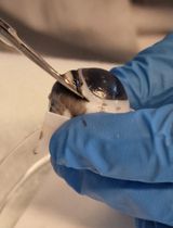
- Harvesting of ovaries from female Xenopus laevis
- Oocyte isolation and maintenance
- Dumont size 4 forceps (Fine Science Tools, catalog number: 11241-30 )
- Clay AdamsTM Nutator mixer (BD, catalog number: 421105 )
- Oocytes transferring pipette: a polished glass Pasteur pipette (Fisher Scientific, FisherbrandTM, catalog number: 13-678-20 )
- Pipette pump (SP Scienceware - Bel-Art Products - H-B Instrument, catalog number: F378980000 )
- B.O.D. low temperature refrigerated incubator (VWR)
- Dumont size 4 forceps (Fine Science Tools, catalog number: 11241-30 )
- Oocyte injection
- Programmable nanoliter injector (Drummond Scientific, model: Nanoject III )
- Steel base plate (WPI, catalog number: 5052 )
- Magnetic holding stand (WPI, catalog number: M10 )
- Three-axis manual micromanipulator (WPI, catalog number: M3301 )
- Stereo microscope (Olympus, model: SZ61 )
- Fiber optic illuminator and bifurcated light guide (WPI)
- Micropipette pullers (NARISHIGE, model: PC10 )
- Programmable nanoliter injector (Drummond Scientific, model: Nanoject III )
- Two-electrode voltage clamp
- Steel base plate (WPI, catalog number: 5479 )
- Steel base plate (WPI, catalog number: 5052 )
- Magnetic holding devices, three (WPI, catalog number: M10 )
- Three-axis manual micromanipulators, three (WPI, catalog number: M3301 )
- Two-electrode voltage clamp amplifier (Molecular Devices, Axon Accessories, model: GeneClamp 500B )
- Headstages (Molecular Devices, Axon Accessories, model: HS-2A )
- Virtual ground bath clamp (Molecular Devices, Axon Accessories, model: VG-2A )
- Pipette holders (Molecular Devices, model: HL-U )
- Ag/AgCl pellets (2 mm diameter, 4 mm long) (WPI, catalog number: EP2 )
- Digitizer (Molecular Devices, model: Digidata 1550B )
- BNC cables (A-M systems)
- Perfusion system (Warner Instruments, model: VC-6 )
- Flow valve (Warner Instruments, model: FR-50 )
- Vacuum attached waste bottle (Fisher Scientific, FisherbrandTM, catalog number: FB3002000 )
- Oocyte chamber (20 mm diameter and 6 mm deep ring with glued glass bottom)
- Silver wire AWG 26 for electrodes (WPI, catalog number: AGW1510 )
- PC computer with Windows operating system
- Steel base plate (WPI, catalog number: 5479 )
Software
- pClamp 10 software (Molecular Devices)
Procedure
文章信息
版权信息
© 2017 The Authors; exclusive licensee Bio-protocol LLC.
如何引用
Readers should cite both the Bio-protocol article and the original research article where this protocol was used:
- Shi, S. and Carattino, M. D. (2017). Expression and Analysis of Flow-regulated Ion Channels in Xenopus Oocytes. Bio-protocol 7(8): e2224. DOI: 10.21769/BioProtoc.2224.
- Shi, S., Luke, C. J., Miedel, M. T., Silverman, G. A. and Kleyman, T. R. (2016). Activation of the Caenorhabditis elegans degenerin channel by shear stress requires the MEC-10 subunit. J Biol Chem 291(27): 14012-14022.
分类
分子生物学 > 蛋白质 > 离子通道信号转导
细胞生物学 > 细胞分离和培养 > 细胞分离
您对这篇实验方法有问题吗?
在此处发布您的问题,我们将邀请本文作者来回答。同时,我们会将您的问题发布到Bio-protocol Exchange,以便寻求社区成员的帮助。