

Assessment of Murine Retinal Function by Electroretinography
通过视网膜电图评估小鼠视网膜功能
发布: 2017年04月05日第7卷第7期 DOI: 10.21769/BioProtoc.2218 浏览次数: 17759
评审: Jyotiska ChaudhuriPascal Fossat Clara Lubeseder-Martellato
参见作者原研究论文
The authors used this protocol in:

Dec 2015


相关实验方案
Abstract
The electroretinogram (ERG) is a sensitive and noninvasive method for testing retinal function. In this protocol, we describe a method for performing ERGs in mice. Contact lenses on the mouse cornea measure the electrical response to a light stimulus of photoreceptors and downstream retinal cells, and the collected data are analyzed to evaluate retinal function.
Keywords: Electroretinogram (ERG) (视网膜电图(ERG))Background
Electroretinograms (ERGs) are used by researchers and clinicians to test retinal function by measuring the electrical response of retinal cells to a light stimulus. The ERG is a useful tool for measuring retinal responses in mice due to its high level of sensitivity and noninvasive nature, and can be utilized to assess eye disease and retinal degeneration (Duncan et al., 2003; Phillips et al., 2010; Zhao et al., 2011; Vollrath et al., 2015). In mouse genetic retinal disease models, ERGs can be used to assess retinal degeneration at multiple time points as the disease progresses (Duncan et al., 2003). For studies evaluating the effect of drug treatment on the mouse eye, retinal function can be assessed before and after treatment in the same eye (Zhao et al., 2011). In the following protocol we describe a method for measuring the functional response of photoreceptors and downstream retinal cells in mice that builds on a previously published approach (Phillips et al., 2010). The protocol can be readily applied to animals three weeks of age and older.
ERGs can be used to measure the electrical response to light flashes in either dark-adapted (scotopic) or light-adapted (photopic) mice. In scotopic ERGs, mice are presented with low intensity light flashes to induce rod activation, so the function of rod photoreceptor and downstream retinal cells can be examined (Fu, 2010). A prolonged period of dark-adaptation is critical to achieve maximal rod sensitivity, and to keep cone stimulation minimal (Pepperberg, 2003). In photopic ERGs, mice are presented with high intensity light flashes after a period of light stimulation. Under these conditions, there is high cone activation and the rod response is suppressed. Therefore, photopic ERGs can be used to measure the function of cone photoreceptors and downstream retinal cells (Fu, 2010).
This method of performing ERGs allows for the calculation of amplitude and time-to-peak of two major waves, the a-wave and b-wave. The a-wave is a measure of the initial response of photoreceptors to a brief flash of light (Brown, 1968; Perlman, 2015). The b-wave is a measure of the response of downstream retinal neurons, including bipolar cells, to photoreceptor stimulation (Brown, 1968; Perlman, 2015). Loss of amplitude in either the a-wave or b-wave may be attributed to a number of retinal dystrophies (Creel, 2015), while ‘supernormal’ waves with increased amplitude have been attributed to cone dystrophies (Phillips et al., 2010).
Materials and Reagents
- Absorbent pads (Bound Tree Medical, catalog number: 111-16650 )
- Syringes and needles (BD, catalog number: 329461 )
- Spray/squirt water bottle (Qorpak, catalog number: PLC-03431 )
- Cotton swabs (Uline, catalog number: S18985PK )
- Two 29 gauge, 12 mm needle electrodes for ground and reference (The Electrode Store, catalog number: GRD-SAF )
- Mice
- Ketamine hydrochloride (NADA: 045-290)/xylazine hydrochloride (NADA: 139-236) cocktail (80 mg/kg/13 mg/kg)
- 1% atropine sulfate ophthalmic solution (NDC: 24208-750-60)
- 2.5% phenylephrine hydrochloride ophthalmic solution (NDC: 17478-200-12)
- 0.5% proparacaine hydrochloride (NDC: 24208-730-06)
- Hydrating eye ointment (Refresh Tears; Allergan, NDC: 0023-0798)
- 2.5% hypromellose (NDC: 17238-610-15)
- 70% ethanol
Equipment
- Dark room
- Red filter (ROSCO, catalog number: Roscolux Supergel R27 Medium Red)
- Black electrical tape (3M, catalog number: 06132 )
- Surgical tape (3M, catalog number: 15270 )
- Ganzfeld ColorDome (Diagnosys, catalog number: D125 ) or similar
- Espion visual electrophysiology system (Diagnosys, catalog number: D315 ) or similar
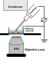
- Two Bayer-Mittag contact lens electrodes (Mt Sinai, Phil Cook) (Bayer et al., 2001)
- Forceps (Fine Scientific Tools, catalog number: 11295-00 )
- Elevated mouse platform (Foam or plastic can be used to fit space and size requirements)
- Non-electric heating pad (Braintree Scientific, catalog number: DPIP )
- Clean mouse cage
- Optional: opaque box (Rubbermaid Commercial Products, catalog number: 9S31 ), blackout cloth (Thorlabs, catalog number: BK5 ), red light headlamp (Princeton Tec, model: Sync headlamp ), red light desk lamp (AstroGizmos, catalog number: DSKLMP22 ), lux meter (Fisher Scientific, catalog number: 06-662-63 )
Software
- Data analysis software such as Microsoft Excel (v 14.7.1) or GraphPad Prism (v 7.0)
Procedure
文章信息
版权信息
© 2017 The Authors; exclusive licensee Bio-protocol LLC.
如何引用
Benchorin, G., Calton, M. A., Beaulieu, M. O. and Vollrath, D. (2017). Assessment of Murine Retinal Function by Electroretinography. Bio-protocol 7(7): e2218. DOI: 10.21769/BioProtoc.2218.
分类
发育生物学 > 细胞信号传导 > 电反应
您对这篇实验方法有问题吗?
在此处发布您的问题,我们将邀请本文作者来回答。同时,我们会将您的问题发布到Bio-protocol Exchange,以便寻求社区成员的帮助。