

In vitro Detection of Neutrophil Traps and Post-attack Cell Wall Changes in Candida Hyphae
念珠菌菌丝中嗜中性粒细胞诱捕和攻击后细胞壁变化的体外检测

发布: 2017年04月05日第7卷第7期 DOI: 10.21769/BioProtoc.2213 浏览次数: 10909
评审: Alka MehraSaskia F. ErttmannSadri Znaidi
参见作者原研究论文
The authors used this protocol in:

May 2016
Advertisement


Abstract
In this protocol we describe how to visualize neutrophil extracellular traps (NETs) and fungal cell wall changes in the context of the coculture of mouse neutrophils with fungal hyphae of Candida albicans. These protocols are easily adjusted to test a wide array of hypotheses related to the impact of immune cells on fungi and the cell wall, making them promising tools for exploring host-pathogen interactions during fungal infection.
Keywords: Fungi (真菌)Background
C. albicans is a polymorphic opportunistic yeast and neutrophils are immune cells critical for defense against this and other fungal pathogens (Brown et al., 2012; Lionakis and Netea, 2013). NETs are a potential defense mechanism that can be deployed against pathogens and it has been suggested that they are preferentially deployed against microbial cells such as C. albicans hyphae that are too large to phagocytose (Urban et al., 2006; Bruns et al., 2010; Branzk et al., 2014; Rohm et al., 2014). NETs have been shown to contain a number of components including myeloperoxidase, extracellular DNA and citrullinated histones (Amulic et al., 2012; Branzk and Papayannopoulos, 2013). For positive identification of NETs, the standard in both in vitro and in vivo experiments includes staining for and demonstrating the colocalization of these markers. While the exact contribution of NETs to defense against C. albicans infection is not well understood, our group has demonstrated they can provoke stress responses and cell wall rearrangement in C. albicans hyphae. Specifically, NET attack results in greater chitin deposition and β-glucan exposure as shown schematically in Figure 1. These polysaccharides normally lie underneath the mannan layer, and their exposure can change immune recognition (Perez-Garcia et al., 2011). The basic assays described here were used extensively to probe this subject by our group (Hopke et al., 2016). While outlined here for the purpose of detecting NETs and fungal cell wall changes, this protocol is easily tweaked to leverage many combinations of chemical inhibitors, transgenic or knockout fungal strains or mouse neutrophils and other staining targets to test a wide array of hypotheses (Hopke et al., 2016). This protocol therefore represents a promising method to further elucidate the impact immune cells have on the C. albicans cell wall, its stress response and the importance of altered epitope exposure to host defense against fungal infection.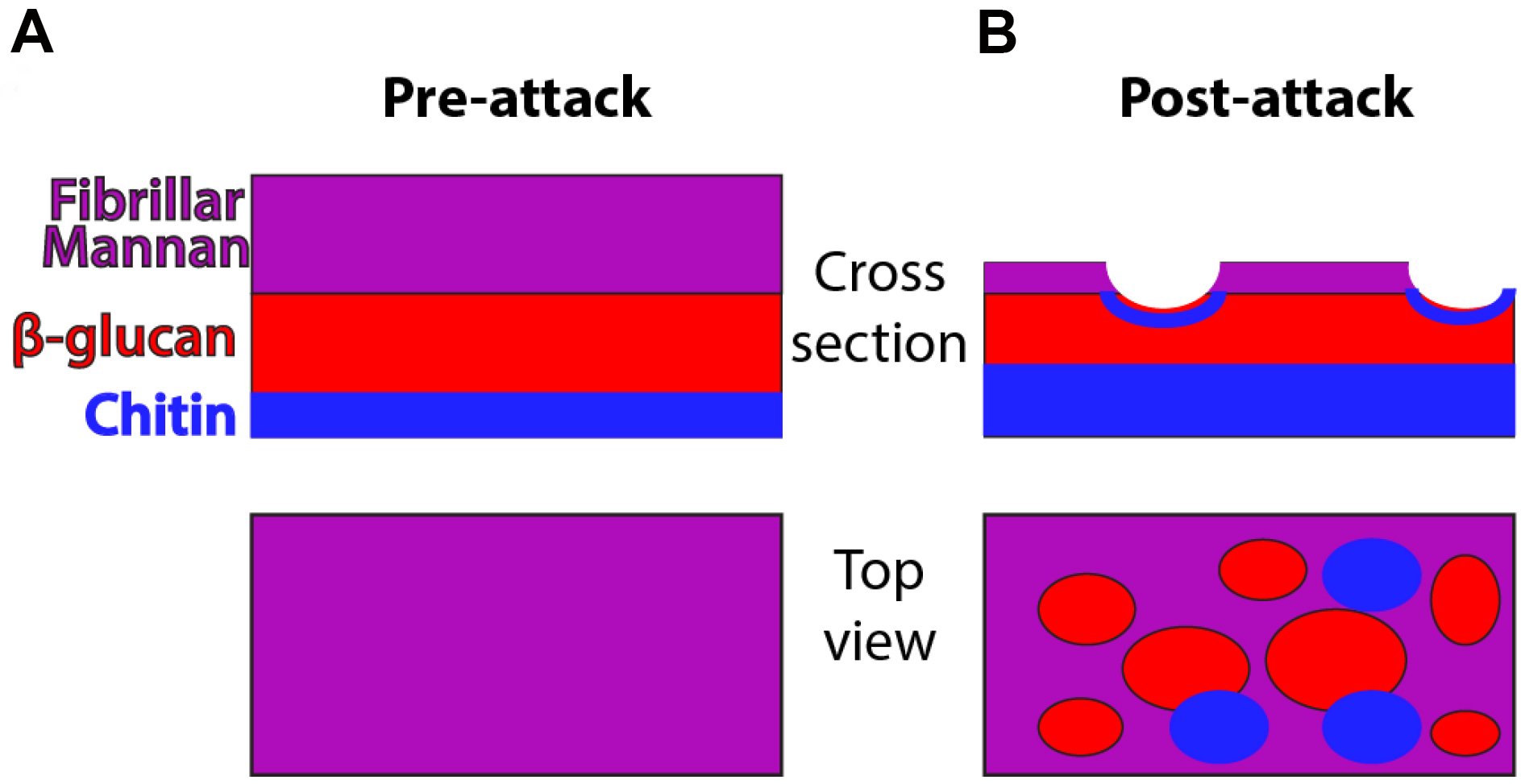
Figure 1. Schematic of cell wall organization pre- and post-neutrophil attack. Under homeostatic conditions, hyphal cell wall is composed of three main polysaccharide components (mannan, β-glucan and chitin) but most chitin and β-glucan are inaccessible for recognition because it lies beneath the mannan layer. Post-attack there is loss of cell wall mannoprotein and increased levels of chitin. These changes lead to greater surface recognition of β-glucan and perhaps also chitin.
Materials and Reagents
- Protective gloves and lab coat
- Flint glass culture tubes 16 x 150 mm (VWR, catalog number: 60825-435 )
- 50 ml conical tubes (Corning, Falcon®, catalog number: 352098 )
- Fisherbrand 100 x 20 mm Petri dishes (Fisher Scientific, catalog number: FB0875711Z )
- 10 ml syringe (BD, catalog number: 301604 )
- 25 G 5/8 needle (BD, catalog number: 305122 )
- 70 µm cell strainers (Corning, Falcon®, catalog number: 352350 )
- 1.7 ml microcentrifuge tubes
- Plain microscope slides (VWR, catalog number: 48300-025 )
- Kim-KapTM Disposable closures; 16 mm (Kimble Chase Life Science and Research Products, catalog number: 7366316 )
- Miltenyi columns (Miltenyi Biotec, catalog number: 130-021-101 )
- WT-FarRed670 strain of C. albicans (Hopke et al., 2016)
- WT-GFP strain (Wheeler et al., 2008)
- C57BL/6J mice, female 6-10 weeks old (THE JACKSON LABORATORIES, catalog number: 000664 )
- Glycerol (EMD Millipore, catalog number: GX0185-5 )
- Anti-Ly6G biotin antibody (Thermo Fisher Scientific, eBioscience, catalog number: 13-5931-85 )
- Anti-biotin magnetic beads (Miltenyi Biotec, catalog number: 130-090-485 )
- Trypan blue (Lonza, catalog number: 17-942E )
- FluoReporter® Cell-Surface Biotinylation Kit (Thermo Fisher Scientific, Molecular ProbesTM, catalog number: F20650 )
- Alexa Fluor 647 conjugated streptavidin (Jackson ImmunoResearch, catalog number: 016-600-084 )
- Anti-myeloperoxidase (MPO) (R&D Systems, catalog number: AF3667 )
- Anti-histone H3 (citrulline R2+R8+R17) (Abcam, catalog number: ab5103 )
- Sytox Green (Thermo Fisher Scientific, Molecular ProbesTM, catalog number: S7020 )
- Calcofluor white/Fluorescent brightener 28 (Sigma-Aldrich, catalog number: F3543 )
- Donkey anti-goat IgG Cy3 (Jackson ImmunoResearch, catalog number: 705-165-147 )
- Donkey anti-rabbit IgG Cy3 (Jackson ImmunoResearch, catalog number: 711-165-152 )
- sDectin-1-Fc (produced in house according to [Graham et al., 2006])
- Donkey anti-human IgG Cy3 (Jackson ImmunoResearch, catalog number: 709-165-149 )
- Nail polish
- Sodium chloride (NaCl) (Fisher Scientific, catalog number: S271-3 )
- Sodium phosphate, dibasic anhydrous (Na2HPO4) (Fisher Scientific, catalog number: S374-500 )
- Potassium phosphate, monobasic anhydrous (KH2PO4) (Fisher Scientific, catalog number: P285-500 )
- Sodium carbonate (Na2CO3) (Sigma-Aldrich, catalog number: S7795 )
- BD Bacto peptone (BD, BactoTM, catalog number: 211677 )
- BD Bacto yeast extract (BD, BactoTM, catalog number: 212750 )
- Dextrose (Fisher Scientific, catalog number: D-163 )
- BD Bacto agar (BD, BactoTM, catalog number: 214014 )
- Heat inactivated fetal bovine serum (Thermo Fisher Scientific, GibcoTM, catalog number: 10082147 )
- Probumin® bovine serum albumin diagnostic grade powder (EMD Millipore, catalog number: 820451 )
- RPMI, with 25 mM HEPES and L-glutamine (Lonza, catalog number: 12-115F )
- Ammonium chloride (NH4Cl) (EMD Millipore, catalog number: AX12701 )
- Crystal violet (Sigma-Aldrich, catalog number: C0775 )
- Acetic acid (Fisher Scientific, catalog number: A38-212 )
- Tris base (AMRESCO, catalog number: 0497 )
- Hydrochloric acid (HCl) (Fisher Scientific, catalog number: A144-500 )
- PBS, pH 7.2 (see Recipes)
- PBS + Na2CO3, pH 8 (see Recipes)
- YPD broth (see Recipes)
- PBS + 5% FBS (see Recipes)
- PBS + 2% FBS (see Recipes)
- PBS + 2% BSA (see Recipes)
- RPMI + 5% FBS (see Recipes)
- Tris-HCl, pH 8.0 (see Recipes)
- TAC buffer (see Recipes)
- Turks solution (see Recipes)
Equipment
- Roller drum (Eppendorf, New Brunswick Scientific) and incubators (VWR)
- Hemacytometer for cell counting (Hausser Scientific, catalog number: 1492 )
- Spectrophotometer (Eppendorf; Biophotometer)
- Centrifuge with adaptors for 15 and 50 ml conical tubes (Thermo Fisher Scientific, model: SorvallTM LegendTM RT )
- AutoMACs separation system (Miltenyi Biotec, model: autoMACS® Pro Separator )
- Zeiss Axiovision fluorescence microscope (Zeiss; custom build)
- Dissection kit (VWR, catalog number: 631-0616 )
- pH meter (Fisher Scientific, model: accumetTM Excel XL15 )
- Microcentrifuge for 1.7 ml tubes (Eppendorf, model: 5424 )
- Autoclave
- 500 ml bottle (VWR, catalog number: 89000-233 )
- Water bath
Software
- AxioVision Rel48 software (Zeiss: https://www.zeiss.com/microscopy/us/downloads/axiovision-downloads.html)
- Photoshop (Adobe Systems, San Jose, CA)
Procedure
文章信息
版权信息
© 2017 The Authors; exclusive licensee Bio-protocol LLC.
如何引用
Readers should cite both the Bio-protocol article and the original research article where this protocol was used:
- Hopke, A. and Wheeler, R. T. (2017). In vitro Detection of Neutrophil Traps and Post-attack Cell Wall Changes in Candida Hyphae. Bio-protocol 7(7): e2213. DOI: 10.21769/BioProtoc.2213.
- Hopke, A., Nicke, N., Hidu, E. E., Degani, G., Popolo, L. and Wheeler, R. T. (2016). Neutrophil attack triggers extracellular trap-dependent candida cell wall remodeling and altered immune recognition. PLoS Pathog 12(5): e1005644.
分类
免疫学 > 免疫细胞成像 > 共聚焦显微镜技术
微生物学 > 微生物-宿主相互作用 > 真菌
细胞生物学 > 细胞染色 > 细胞壁
您对这篇实验方法有问题吗?
在此处发布您的问题,我们将邀请本文作者来回答。同时,我们会将您的问题发布到Bio-protocol Exchange,以便寻求社区成员的帮助。